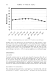
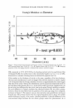
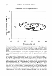
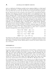
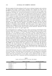
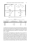
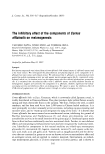
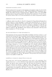
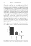
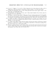
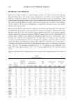
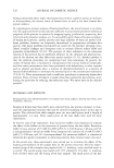
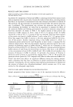
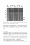
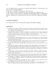
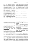
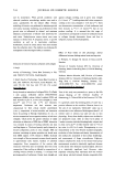
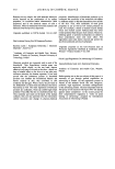
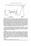
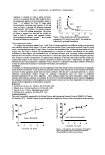
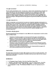
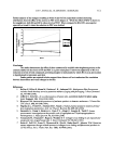
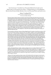
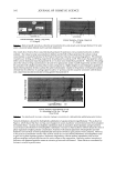
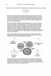
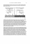
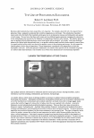
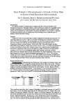
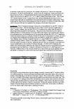
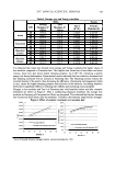
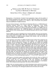
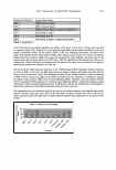
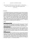
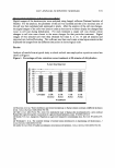
586 JOURNAL OF COSMETIC SCIENCE INFRARED MICRO-SPECTROSCOPIC IMAGING OF CHANGES IN NATURAL MOISTURIZING f AClOR (NMF) IN HUMAN STRATUM CORNEUM David J. Moore, Ph.D. International Specialty Products, Global R&D, Wayne, NJ, USA Introduction Recent advances in infrared (IR) spectroscopic imaging and confocal Raman micro-spectroscopy have permitted the acquisition of spatially resolved chemical composition and structural information from biologically important samples [1,2). Challenges in data collection and interpretation are being confronted and SUIInOunted as the work. moves from serving as an adjunct to histopathology to playing a more predictive role in diagnosing disease, providing insights into the molecular interactions preceding the onset of disease, and tracking the effectiveness of therapeutics on a molecular level. In the current study, the maturation process of individual human Optical and lnl'rared lma&lng or hmnan comeocyta. Representative IR 1pectn or comeocytn Isolated 1n,m the 11th (bottom spedrum) md 3rd (top spectrum) tape strips. comeocytes obtained from different depths in the SC was investigated using IR micro--spectroscopic imaging. Instrumental details and experimental methods have been described [ 4,5). The results from experiments conducted on cells isolated from healthy skin reveal depth dependent compositional differences and provide an initial baseline from which an examination of the variability between subjects, body sites, age, and a variety of disease states can be made. Our recent studies indicate that changes in NMF levels induced by skin cleansing can be directly detected via IR micro spectroscopic imaging of corneocyles from washed versus control sites at the same depth within the stratum comeum. Materials and Methods Corneocytes from different SC layers were collected from human foreann skin by sequential tape stripping. The first two tape strips were discarded and comeocytes collected from the third, sixth, and eleventh tape strips. IR microscopic images were acquin:d with the Perk.in-Ebner Spotlight system consisting of an essentially linear array (16 x I detector elements) mercury cadmiwn-telluride (Men. About SO comeocytes from each layer were randomly selected and IR images (60 x 60 microns) were acquired with a pixel size of 6.2S microns. Each comeocyte image was acquin:d in approximately 20 minutes. ISys 3.0 software (Spectral Dimensions, Maryland) was used for analysis and construction of the IR images. Linear baselines were applied A I•··:_ - /\ 1� � --v � \ Jij/ "'-.__/_\... .� I N� - '- UH UH 1111 UH HH � Wavenumberlem "' Mean Infrared lm1111Dg spectn of multiple comeocytes from two depths In human SC compared to IR spectnim or model NMF mm. over spectral regions of interest Data interpretation was facilitated by averaging the spectra of comeocytes from particular stratwn comewn layers to improve signal to noise ratios. Correlation coefficients were then calculated between the mean spectrum of each layer and each spectrwn contained in an image to generate correlation maps.

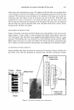
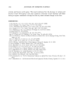
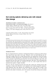
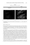
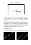
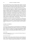
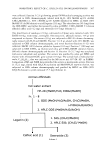
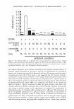
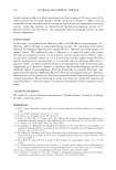
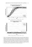
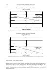
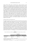
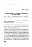
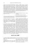
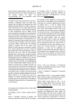
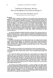
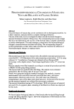
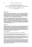
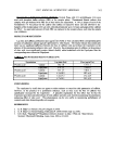
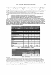
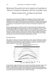
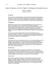
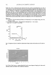
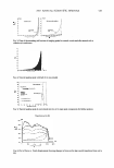
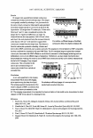
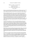
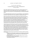
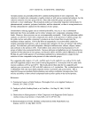
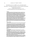
2007 ANNUAL SCIENTIFIC SEMINAR Results IR images were acquired from multiple comeocytes isolated from the third and eleventh tape strips. The images were spatially masked by selecting a 4 x 4 pixel area from the center of each comeocyte. Mean spectra wen: generated from these multiple spectra of comoocytes as shown in the figure below. IR images from 72 comcocytes, 36 from each from layer 3 and 11, were concatenated to produce the images sho\W. Significant differences can clearly be observed between the mean spectra (1180-1430 cm- 1 ) from each layer, the most prominent being the incr� intensity a,r. .... ,.._.. _ _........_ :I 587 • in the band at 1404 cm·1 in the spectrum acquired from Correlation coefficient images of multiple comeocytes isolated from the deeper layer. This feature is comeocytes from two depths in human SC. from the carboxylate symmetric stretching vibration and derives from NMF constituents, such as amino acid salts. The assignment of these features to NMF components has been confirmed by comparison with pure NMF films. The IR correlation images acquired from isolated comeocytes demonstrate that NMF diffen:nccs can be directly measured via this imaging technology and can diffcrc:ntiate comeocytes at different stages of maturation. Furthermore, our most recc:nt ccpcriments have demonstrated that washing skin results in loss of NMF from comeocytes and this can be directly measured and tracked with IR imaging of tape stripped comeocytes. This is illustrated in the spectra and correlation images of comeocytcs isolated form washed and wtwashed skin. Conclusion A new !Cllli-quantitative skin imaging measurement which utilizes IR imaging Ill 1n1..-ofan-,.6---• lllduw.-.dma micro-spectroscopy has been developed. Correlation coefficient imqes of comeocytes from The technique permits measurement of the w■s•ed and unwashed skin sites. relative changes in NMF concentration that occurs with comeocyte maturation in the SC. This imaging measurement of an important biochemical marker of skin health is also demonstrated to detect changes in NMF levels induced by cleansing the skin. Reference 1. Boskey AL, Moore DJ,Amling M, Canalis E, Delany AM Jou ma I of Bone and Mineral Research 18(6):1005-1011, 2003 2. Mc:ndelsohn R, Chen H-C, Rerek ME, Moore DJ. Joumal of Biomedical Optics 8(2): 185-190, 2003 3. Xiao C, Moore DJ, Rerek ME, Flach CR, Mendelsohn R. Journal oflnvutigative Dermatology 114:622- 632, 200S 4. Xiao C, Moore DJ, Flach CR, Mendelsohn R. Vibrational Spectroscopy38:1SI-158, 2005 5. Zhang G, Moore DJ, Mendelsohn R, Flach CR. Journal ofln11estigative Dermatology 126: 1088-1094, 2006
Purchased for the exclusive use of nofirst nolast (unknown)
From: SCC Media Library & Resource Center (library.scconline.org)