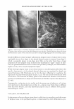
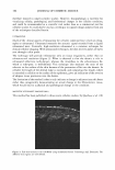
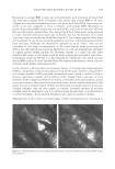
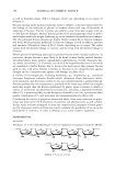
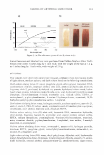
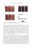
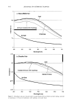
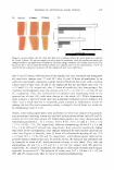
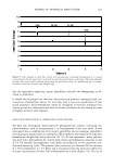
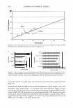
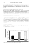
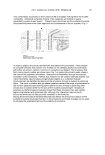
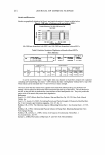
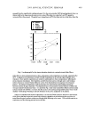
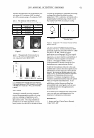
390 JOURNAL OF COSMETIC SCIENCE rule out the influence of long-term sun exposure as compared to actinic damage (62). However, the limitation of this technique, as with other image analysis systems, is in obtaining clear resolution beyond the upper portion of the dermal papillae. CONTACT THERMOGRAPHY Contact thermography (CT) can measure alterations or variations in skin temperature, and thus any skin disorder affecting directly or indirectly the microcirculation of the skin becomes a candidate for thermal evaluation. Consideration of the application of CT to cellulite should be given. In a compressed subcutis, as seen in cellulite and visualized by several of the image techniques described in this paper, the microvasculature is pressed against the papillary dermis. The amount of heat generated could be compared to that observed in non-cellulite skin. CONCLUSIONS Dimpling of the skin of the peripheral regions of the female body, commonly called cellulite, is presumed wrongly to be an abnormality of adipose tissues rather than normal reality. Reality dictates that cellulite is a condition reflected in differences in adipose biochemistry and connective tissue structure in the female gluteal-femoral adipose tis sue. At this stage of current understanding of the condition, it is concluded that "visual cellulite" is a consequence of the inability of collagen at the papillary dermis to contain adipose protrusion through a "thin" dermis, which is not observed in males. From current image analysis techniques, it is suggested that dimpling of the skin is associated with enlarged fat lobules surrounded by thin and focally loose connective tissue strands. Furthermore, the "striae" observed in these studies may result from excessive tension arising from the continuous and progressive vertically orientated stretch in the adipose tissue. Due to the limitations of each technique, there is no one method at present that can be used to evaluate cellulite. Furthermore, current formulations do not penetrate suffi ciently into the adipose tissue to cause an effect. However, strengthening a "thin" dermis would be a step in the right direction. Although cellulite appears incurable, the subject itself needs a clearer understanding, a consistent approach, coherency to prevent misinterpretation and misunderstanding in both the scientific and popular literature, formulations that "deliver," correlation of data, good clinical development of methods, and management and understanding of the psychological and social aspects of the condition. Only then can the unmet need of managing the expectations of the consumer and of the regulatory and advertising au thorities be achieved. ACKNOWLEDGMENTS The authors thank Professor Gerald Pierard for permission to use the photographs m Figure 1, and Lucid Inc. for performing the confocal microscopy analyses.

ANALYSIS AND REVIEW OF CELLULITE 391 REFERENCES (1) www.wpatten.clara.net/fat.html. Has Narcissism become 'a metaphor for the human condition?' (April 2004). (2) F. C. Wulf, J. Sandby-Moller, T. Kobayasi, and R. Gnaidecki, Skin ageing and natural photoprotec tion, Micron., 55, 185-191 (2004). (3) M. Tzaphlidou, The role of collagen and elastin in aged skin: An image processing approach, Micron., 35, 173-177 (2004). (4) J. Labat-Robert, Age dependent remodeling of connective tissues: Role of fibronectin and laminin, Pathol. Biol. (Paris), 51, 563-568 (2003). (5) C. Djurhuus, C. Gravholr, S. Nielsen, et al., Additive effects of cortisol and growth hormone on regional and systemic lipolysis in humans, Am.]. Physiol. Endocrin. Metab., 286, 488-494 (2004). (6) A. Peter, U. Schweiger, L. Pellerin, et al., The selfish brain: Competition for energy resources, Neurosci. Biobehav. Rev., 28, 143-180 (2004). (7) F. Frey, A. Odermatt, and B. Frey, Glucocorticoid-mediated mineralocorticoid receptor activation and hypertension, Curr. Opin. Nephrol. Hypertens., 13, 451-458 (2004). (8) G. E. Pierard, J. L. Nizet, and C. Pierard-Franchimont, Cellulite: From standing fat herniation to hypodermal stretch marks, Am. J. Dermatopathol. 22, 34-3 7 (2000). (9) F. Mirrashed, J. Sharp, V. Krause, et al., Pilot study of dermal and subcutaneous fat structures by MRI in individuals who differ in gender, BMI and cellulite grading, J. Skin Res. Technol., 10, 161-168 (2004). (10) B. Querleux, C. Cornillon, 0. Jolivet, et al., Anatomy and physiology of subcutaneous adipose tissue by in vivo magnetic resonance imaging and spectroscopy: Relationship with sex and presence of cellulite, J. Skin Res. Technol., 8, 118-124 (2002). (11) T. Lotti, I. Ghersetich, C. Grappone, and G. Dini, Proteoglycans in so-called cellulite, Intl. J. Der matol., 29, 272-274 (1990). (12) C. Bertin, H. Zunino, J.C. Pittet, et al., A double-blind evaluation of the activity of an anti-cellulite product containing retinol, caffeine and ruscogenine by a combination of several non-invasive methods. ]. Cosmet. Sci. 52, 199-210 (2001). (13) C. Pierard-Franchimont, G. E. Pierard, F. Henry, V. Vroome, and G. Cauwenbergh, A randomized placebo-controlled trial of topical retinol in the treatment of cellulite, Am.]. Dermatol., l, 369-374 (2000). (14) F. Nurnberger and G. J. Muller, So-called cellulite: An invented disease, Dermatol. Surg. Oncol., 4, 221-229 (1978). (15) S. B. Curri, Cellulite and fatty tissue microcirculation, Cosmet. Toiletr., 108, 51-58 (1993). (16) S. B. Cutri, and E. Bombardelli, Local lipodystrophy and districtual microcirculation, Cosmet. Toiletr., 109, 51-65 (1994). (17) Z. D. Draelos and K. D. Marenus, Cellulite etiology and purported treatment, Dermatol. Surg., 23, 1177-1181 (1997). (18) M. Rosenbaum, V. Prieto, J. Hellmer, et al. An exploratory investigation of the morphology and biochemistry of cellulite, Plast. Reconstr. Surg., 101, 1934-1939 (1998). (19) A. M. Kligman, Cellulite: Facts and fiction,]. Geriatr. Dermatol., 5, 136-139 (1999). (20) P. Bjorntorp, M. Karlsson, and P. Pettersson, Expansion of adipose storage capacity at different ages in rats, Metabolism, 31, 366-3 7 3 (1982). (21) M. Rebuffe-Scrive, M. Bronnegard, A. Nilsson, et al., Steroid hormone receptors in human adipose tissue,]. Clin. Endocrin. Metab., 71, 1215-1219 (1990). (22) P. Bjorntorp, Adipose tissue distribution and function, Int.]. Obesity 15, 67-81 (1991), and references cited therein. (23) P. McTernan, L. Andersen, A. Anwar, et al., Glucocorticoid regulation of P450 aromastase activity in human adipose tissue: Gender and site differences,]. Clin. Endocrinol. Metab., 87, 1327-1338 (2002). (24) S. Boullu-Ciocca, 0. Paulmyer-Lacroix, F. Fina, et al., Expression of the mRNA's codifying for the glucocorticoid receptor isoforms in obesity, Obesity Res., 11, 925-929 (2003). (25) C. Gravholt, R. Dall, J. Christiansen, et al., Preferential stimulation of abdominal subcutaneous lipolysis after prednelisone exposure in humans, Obesity Res., 10, 774-811 (2002). (26) J. Katz, V. Mohamed-Ali, P. Wood, et al., An in vitro study of the cortisol-cortisone shuttle in subcutaneous abdominal tissue, Clin. Endocrinol., 50, 63-68 (1999). (27) S. Petersen, K. Kristensen, and B. Richelson, Anti-glucocorticoid effects of progesterone in vivo on rat adipose tissue metabolism, Steroids., 68, 543-550 (2003).
Purchased for the exclusive use of nofirst nolast (unknown)
From: SCC Media Library & Resource Center (library.scconline.org)