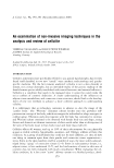
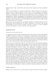
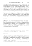
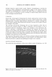
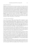
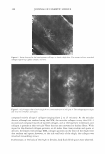
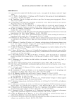
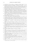
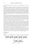
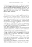
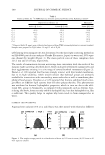
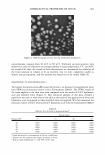
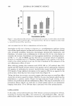
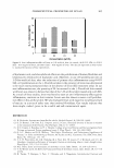
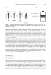
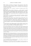
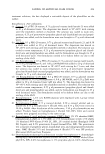
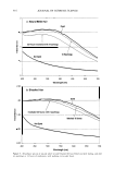
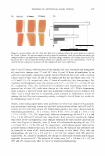
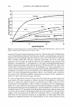
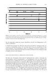
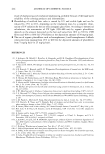
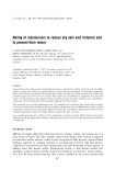
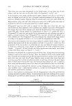
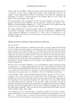
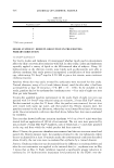
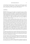
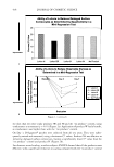
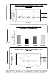
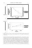
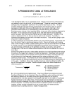
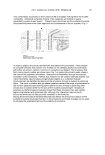
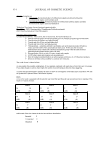
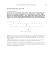
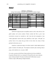
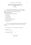
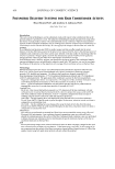
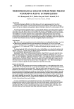
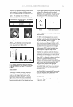
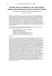
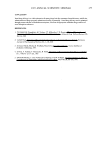
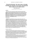
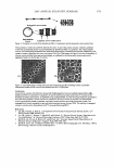
388 JOURNAL OF COSMETIC SCIENCE Figure 4. Striae observed at the basal-spinous cell layer in female thigh skin. The arrows indicate stretched collagen appearing tightly compact and taut. Figure 5. (a,b) Images taken of male thigh skin at same resolution as in Figure 4. The collagen appears light and very fine ("fluffy") and sparse. composed mainly of type I collagen (ranging from 2 to 15 microns). At the reticular dermis, although not resolved using the CFM, the reticular collagen is very thin (0.2-1 micron) and composed mainly of type III collagen, and at the basement membrane zone collagen is present as thin reticular fibers that are not converted to thicker fibers. With regard to the observed collagen patterns, in all males these were random and sparse at all sites. In females with average BMI, collagen patterns on the front of the thigh were also random and sparse however, at the side and back of the thigh, the collagen was stretched (resembling striae). Furthermore, at the back of the thigh in females, dark fluid-filled spaces were observed.

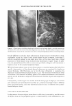
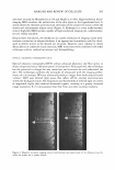
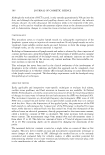
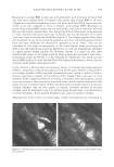
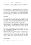
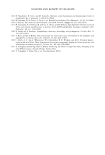
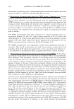
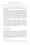
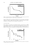
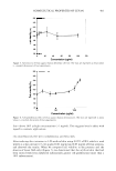
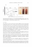
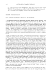
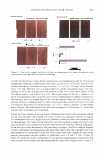
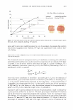
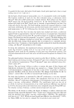
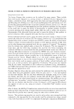
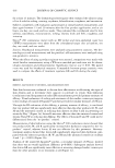
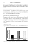
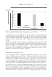
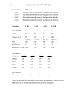
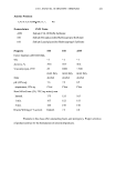
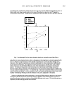
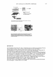
ANALYSIS AND REVIEW OF CELLULITE 389 Resolution in average-BM! women was still permissible with resolution of blood flow and individual collagen fibers. In females with greater than average BMI at all sites, collagen was stretched (resembling striae), and many dark fluid-filled spaces were ob served at all sites compared to those in females with average BMI. Resolution in greater-than-average-BM! women was not permissible, with loss of resolution of blood flow and individual collagen fibers. The observed dark fluid-filled spaces index-matched to water. Another observation particular to female skin was the presence of so-called "puncture" zones surrounded by blood flow (Figure 6). The collagen appeared very dense and "stretched" in one direction and the epidermis very thin, with little resolution of the basal cell layer. Although the observations appeared as actual "holes," they are not considered at this stage of investigation to be actual adipose tissue puncturing the dermis, but the indentations as seen by Querleux et al. (10) and subsequently identified as tightly packed dermal papillae (9). However, neither is it ruled out that these observed dark spaces in the image could be where adipose had protruded, since histo pathological studies by Pierard et al. had observed fat "herniation" (8), which was also seen in MRI studies (9). Since the dark fluid-filled spaces index match to water, they are currently under further investigation (62). In the cellulite condition there are numerous reports of increased glycosaminoglycans (GAGs), components of the ground substance that fill the spaces between collagen fibers and collagen bundles. GAGs trap bulk (tetrahedron) water, which is unable to bind to proteins, resulting in rigidity and instability of the collagen fibers-also seen in striae distensae. If the collagen as observed in cellulite is stretched, more spaces appear, giving rise to an increase in the potential for more bulk water, which could be observed as dark spaces that are not blood vessels. If there are abnormally increased amounts of reticular collagen (thinner), they are then suspect to rupture. Increased amounts of reticular collagen could be examined to see if in cellulite-prone females there is an abnormality in active fibroblasts. Active growth fibroblasts also cause an increase in GAGs. Although this work is still in its initial stages, further investigations are continuing to Figure 6. (a,b) Indentations into the dermal-epidermal layers. The arrows indicate actual blood flow within the capillaries.
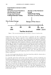
Purchased for the exclusive use of nofirst nolast (unknown)
From: SCC Media Library & Resource Center (library.scconline.org)