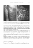
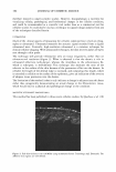
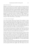
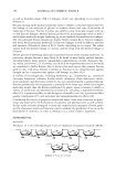
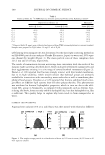
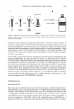
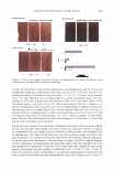
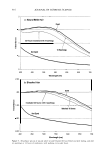
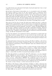
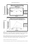
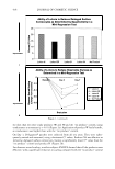
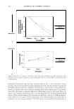
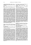
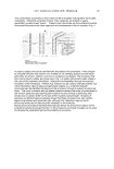
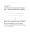
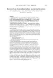
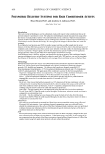
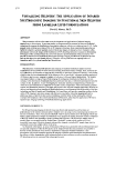
380 JOURNAL OF COSMETIC SCIENCE opment needs to take a wider look at the causes of the condition, not only its manifes tation. There is little coherency or consistency within the scientific literature as to the mani festation of the "cellulite condition" (9-17). From a clinical viewpoint, biochemical markers in adipose tissue metabolism can play a key role in understanding the cellulite condition and, therefore, in determining the effectiveness of an active/product in the management of cellulite. Furthermore, the development of clinical methods, which can correlate in vitro studies, will finally bring coherence and understanding to the scientific literature and to the condition, and will ultimately manage the continued expectations and unmet needs of the consumer. Before examining the available clinical imaging methods for cellulite prognosis and the determination of the effectiveness of cosmetic treatments, an overview of the current understanding (or lack of) of the cellulite condition is required, in terms of adipose structure and influences, and of connective tissue organization. ADIPOSE TISSUE ADIPOCYTE SIZE AND ORGANIZATION It is understood from histological examination that the adipose structure within females is different from that of males. Fat accumulation causes pressure, which results in a "hernia" protruding into the dermis and epidermis (8-10). The observed effect (Figure 1) is of dimpling of the skin, notably on the thighs and buttocks, with histopathological observations clearly highlighting fibrosclerotic tissue structures surrounding the adipose tissue, with resultant striae at the dermal/epidermal interface (8). Adipose tissue is a specialized connective tissue that functions as the major storage site for fat (triglycerides). The major bulk of adipose tissue is a loose association of lipid-filled adipocytes held in a framework of collagen fibers. It also contains stromal, vascular cells including fibroblastic connective tissue cells, leukocytes, macrophages, and pre adipocytes that contribute to structural integrity. Distribution in humans is dependent on genetic and environmental factors. The total and regional masses of adipose tissue are dependent on the number of adipocytes as well as their degree of filling with depot fat. Once new adipocytes are formed, they remain. Increasing numbers of adipocytes have far-reaching consequences for the treatment and prevention of obesity. Currently avail able evidence does not suggest a specific regional regulation of fat cell multiplication in subcutaneous depots, which instead seems to occur to a certain critical degree in the filling of available adipocytes (18). The control of the rate of filling of adipocytes seems to be the main factor in determining the local, regional mass of adipose tissue. The balance between the lipid accumulating and mobilization processes in turn regulates this. The steroid hormones exert major effects on these processes (19,20). It appears likely that the resulting effect of the rate of secretion of various steroid hormones, and the local density of their specific receptors, decides the regional distribution of body fat. GENDER DIFFERENCES Adipose tissue is the source of a critical endocrine signal in the control of body weight.

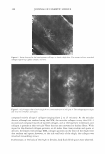
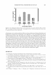
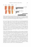
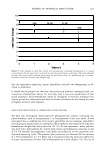
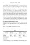
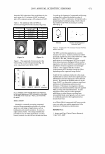
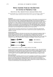
ANALYSIS AND REVIEW OF CELLULITE 381 Figure 1. Clinical aspects of cellulite dimpling (a) of the skin on the female thighs is a visible characteristic of cellulite. Note also the torsion of the skin diagonally across the skin. The full-thickness biopsy (b) shows the fibrosclerotic nature of the connective tissue, with corresponding bulging of the surrounding adipose. Gender differences include a larger subcutaneous adipose tissue in women than in men, explainable in part by a depot in the gluteal-femoral region in women ("pear shape"), which is essentially absent in non-obese men. Men, on the other hand, have a larger proportion of their adipose tissue localized intra-abdominally ("apple" shape). In addi tion, the gluteal-femoral fat cells in women are specifically enlarged, and demonstrate a higher activity of the enzyme lipoprotein lipase. While the larger adipose tissue in non-obese women is considered genetically linked, the specific characteristics of the gluteal-femoral adipocytes are regulated by female sex steroid hormones (20). Hormones are by far the most influential in regulating the deposition of gluteal-femoral adipose tissue (21,22). The sex steroid hormones are never active alone: corticosteroids are always present. The interactions between corticosteroids and sex steroid hormones in adipocyte metabolism are therefore of particular importance (23--43). Consequently, conducting in vitro studies with the wrong type of adipocyte cell culture will clearly lead to misinterpretation of data (i.e., adipose tissue from males is different from that from females, and intra-abdominal adipose tissue is different from gluteal femoral adipose tissue). V ASCULARITY AND INFLAMMATION In the presence of excess adipose tissue there is a deficiency in vascularity, and this excess of adipose tissue in the peripheral regions is characterized by mild inflammation, pro-
Purchased for the exclusive use of nofirst nolast (unknown)
From: SCC Media Library & Resource Center (library.scconline.org)