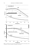
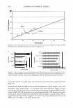
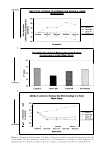
382 JOURNAL OF COSMETIC SCIENCE ducing cytokines, chemokines, and angiogenic factors. It has been suggested that the inflammation is principally an adaptive response to hypoxia in clusters of enlarged adipocytes within an expanding adipose mass (44). With this poor vascularity, there is an increase in the likelihood of edema and venous insufficiency, leading to the phenom enon of "heavy legs" often felt by many women. It could be that since adipose tissue is an active endocrine and paracrine organ that releases a large number of cytokines that influence body weight homeostasis, inflammation, fibrolysis, and insulin resistance, increased levels of saturated fatty acids drive oxidative stress, which in turn increases the level of cytokines/adipokines (45-48). CONNECTIVE TISSUE Studies involving magnetic resonance imaging (MRI) of cellulite have indicated that in females the dermis is thinner than in males (9,10). Females with a high body mass index (BMI) have more cellulite than low BMI women, and high BMI females have less connective tissue in the adipose tissue. Therefore, extrusion is increased and made worse by a thinner dermis. In females cellulite skin has a thinner dermis than non-cellulite skin. In females with cellulite, the amount of protruded fat in the dermis is higher than in males and in females with little or no cellulite. In females with cellulite, there are fewer septa than in males. In females the upper part of subcutaneous tissue is thicker than in males, and the fat chambers are bigger and radial-hence bulging and com pression in the skin above. In males the adipose tissue is supported by "chicken wire," which, with a thicker dermis than in females, prevents gross protrusion into the upper skin. Therefore, the role of supporting connective tissue is important and cannot be dismissed. GLYCOSAMINOGLYCANS Glycosaminoglycans (GAGs) are an integral part of connective tissue structure with high water-attracting properties. There is conflicting evidence in the literature as to the role of GAGs in the cellulite "condition." Lotti et al. (11) argue that an increase in GAGs leads to edema, whereas Querleux et al. (10), through in vivo magnetic resonance im aging, could not confirm this hypothesis neither could Pierard et al. (8). However, the relevance of water "structure" should not be dismissed lightly. The total proportion of water in aging skin increases by 30%, whereas skin hydration (i.e., the amount of water molecules interacting with macromolecules) does not increase. This is because the water is present as bulk or tetrahedron structures, which do not contribute to the stabilization of proteins (49). Increased GAGs trap water, which prevents its access to proteins. Consequently, there is stiffening of collagen and a lack of elasticity in elastin. A recent non-invasive device has been reported (50) for local measurements of changes in tissue water (edema) in human subcutaneous fat in patients undergoing hemodialysis. This technique could find application in cellulite studies (see section on Clinical Con siderations) and in an understanding as to whether "bulk" water is implicated in the manifestation of cellulite. FIBROSCLEROSIS In the cellulite condition, reduction of fat 1s easily achieved by changes in diet and

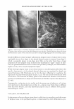
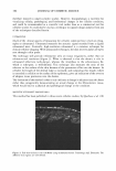
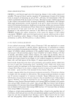
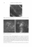
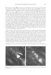
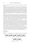
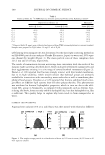
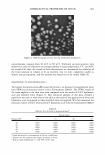
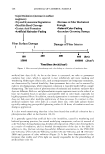
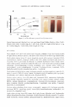
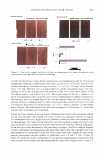
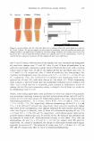
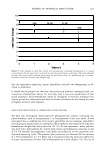
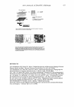
ANALYSIS AND REVIEW OF CELLULITE 383 exercise. However, resolving the fibrosclerotic tissue remains a challenge, as does the increased amount of adipocyte membranous tissue since, although adipocyte content can be reduced, the number of cells is not reduced. Furthermore, understanding why fibro sclerotic tissue forms in this manner needs more in-depth investigation. This can be gleaned from information in studies on breast tissue (51-53) and scar formation and reduction (54,55), as well as from anti-aging studies. As an active endocrine and para crine organ that releases a large number of cytokines that influence inflammation and fibrolysis, increased levels of saturated fatty acids would increase the level of cytokines (adipokines). Consequently, chronic exposure of fibroblasts to cytokines results in fibro sis. Often associated with the cellulite condition are "colorless" striae-striae alba (56), seen as shiny fine lines in the skin. These striae are thought to occur under the pressure of fat accumulation. Seventy percent of females have such striae, including anorexia nervosa patients (12-13 % of the female population). The striae are caused by the presence of more rigid cross-linked collagen, which is easily ruptured under stress. Furthermore, in striae tissue there is an increase in cortisol activity (5 7). LYMPH FLUX Arguments exist in the scientific literature as to the involvement of insufficient lym phatic transport and lymphedema in the cellulite condition, and studies in this area are clearly lacking, giving rise to much controversy (15-17). However, insufficient lym phatic transport in adipose tissue leads to the accumulation of macromolecular proteins and hyaluronan within the extracellular space (58). In lymphedema there is also an accumulation of lymphocytes (macrophages), as well as an increase in the fibroblast, keratinocyte, and adipocyte content of the affected tissue (59). The overgrowth of connective and adipose tissue (also seen in cellulite) in skin and subcutaneous tissue is accompanied by an increase in collagen deposition. EVALUATING THE CONDITION: CLINICAL CONSIDERATIONS There are a number of clinical methods available to evaluate the cellulite condition, though the limitations of each method alone are such that more than one method 1s required in order to correlate clinical findings with any laboratory findings. HISTOP ATHOLOGY The closest work to date as to the real understanding of the cellulite condition is that of Pierard et al. (8). Histopathology tells us that in cellulite superficial fat lobules protrude into the dermis (papillae adiposae), and that there is an unevenness of collagen and elastin fibers. At the site of dimpling, fibrous strands are enlarged, reminiscent of striae distensae (Figure 1). There is also a high content of acid proteoglycans and a2-macroglobulin. There is no tissue dystrophy or lipoatrophy, and the bumpy appear ance is due to a network of connective tissue strands strongly tethering the dermis to the deeper layers, thickened and fibrosclerotic. The limitation of this method is that it is a highly invasive ex vivo technique, and
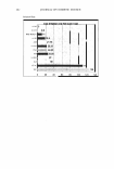
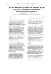
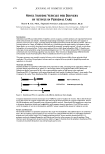
Purchased for the exclusive use of nofirst nolast (unknown)
From: SCC Media Library & Resource Center (library.scconline.org)