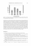
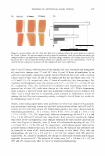
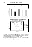
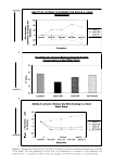
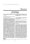
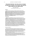
476 JOURNAL OF COSMETIC SCIENCE VISUALIZING DEi.IVERY: THE APPLICATION OF INFARED SPECTROSCOPIC IMAGING TO FUNCTIONAL SKIN DELIVERY FROM lAMEllAR LIPID FORMULATIONS David J. Moore, Ph.D. International Specialty Prodcuts, Wayne, NJ 07470 ABSTRACT The presentation will describe some recent developments and applications of infrared imaging spectroscopy to skin science. In previous srod:ies we demonstrated the potential of this new biomedical technology by imaging .the distribution of penetration enhancers within ex vivo skin sections [ 1,2,3]. In the preserrt work, utilizing newer state-of-the-art IR imaging technology, these experimental approaches are fiiirther developed to generate spatially resolved molecular images of skincare actives. Examples in this presentation will include images of the distribution of UV absorbing molecules in sunscreen formulations and images of the sustained release of actives from lametlar lipid formulations. This presentation will provide an introduction to biomedical infrared spectroscopic imaging and illustrate its potential in areas such as skin deposition and dermal delivery. Examples wil1 be given from our ongoing studies of deposition and delivery ftm:n lamellar lipid formulations. INTRODUCTION The utilization of infrared {IR} spectroscopic imaging to examine biomedical samples has been established in several studies •Of tissue sections inc1uding mineralized tissue, cartilage, brain, and breast [4- 6]. Furthermore, the feasibility ofapplying this technology to questions of physical chemistry, such as drug solubility has also been demonstrated. In our laboratory we are specifically concerned with the application of IR micro-spectroscopic imaging to samples of relevance to skin and topical formulations. The samples investigated address a range of topics from imaging the endogenous distribution of molecules in skin sections, to the distribution of molecules that have penetrated into skin, or delivered and deposited onto the surface of skin. In addition:, we are investigating the molecular dynamics and partitioning of molecules within skin-care formulations as a function of variables such as temperature, water concentration, polymer content, and formulation structure. This paper wi:11 present a few selected examples particularly focusing on delivery and deposition from lamellar gel lipid formulations, as an illustration both of the utility of this new technology and that of lamellar gel formulations. It is hoped these wiH provide compelling. evidence for the utility of IR imaging to provide a unique, and otherwise unavailabie, molecular imagirtg approach to addressing topics of interest in skin science. In keeping with the theme of this meeting the selected examples will emphasize skin delivery and skincare formulations, ra:thet than the application ofIR micro spectroscopic imaging technology to biomedical histology applications. METHODS AND RESULTS For IR spectroscopy imaging of skin the tissue was frozen and mounted to yield sections cut perpendicular to the stratum corrteum surface. Samples were cut using a Leica cryostat giving sections of 5 microns in thickness These were placed on BaF2 IR windows and used directly for lR imaging without any further sample preparation. Sample of formulations were prepared by adding measured volumes of rnaterial to BaF 2 IR Windows and spreading to form a uniform film of product. IR microscopic images were of product films were acquired with the Perkin-Elmer "Spotlight" syste'rn, which consists ofa 16 x 1 essentially linear array of detector elements along with a computer controlled XY sample stage which is automated to move the sample relative to the array. As illustrated in the figures, an array of thousands ofIR spectra is generated with each measurement and any spectral component within the IR spectra can be imaged. From these measurements it is possible to directly see the distribution of any active or formulation component with the formulation. This presentation will provide examples applying IR imaging to lamellar lipid formulations and demonstrated the efficacy of these formulations in the sustained release of active release, as well as excellent retention of UV absorbing actives in sunscreen films submerged in water.

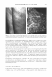
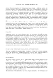
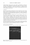
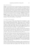
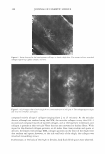
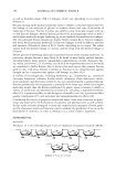
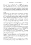
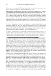
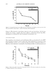
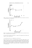
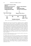
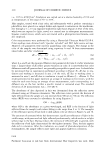
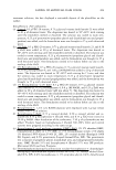
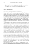
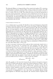
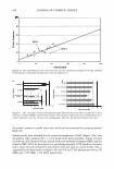
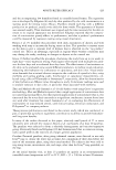
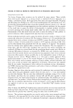
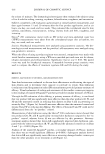
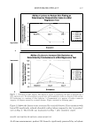
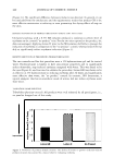
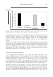
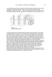
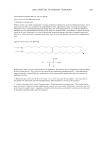
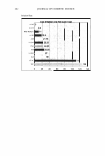
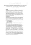
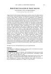
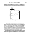
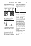
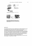
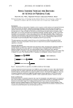
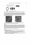
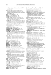
2005 ANNUAL SCIENTIFIC SEMINAR IR array ckttector ! Mkrosc:ope � I Spectrometer I - array ot 4096 complete IR spectra - 1: 1 mapping of 7x7 µm sample spot lo deteclor element Figure 1: Schematic of R imaging m1aoscopewtth6'x&• element anayoetecro, The Ii,.• array s,.tems bulld up a atray of lla1'1 by rnow,g gmple stage - .... Flgulw Z: The opllcal image ol a lamellar lipid sunscreen 111m and the spectrum fntn one elemenl in lhe collected aray ol data. The specsun smws 1he un,que c-;a,o peak ot lhe UV absorbing molecues. The IR spectroscopic Im• ,,_, the rotio ol the UV obsorbin1 sun,c,- loctocryl"""I intensity 111alnst that ol the lip d cti.in, (2218/2852 cm·•]. Mote, the dlfleN!nce in Intensity ocron this lmote does not represenl dlfleence, in film thickneH but rather the mole(ular distribution ol sunscree-n tn the formulMion. REFERENCES 477 [l] R. Mendelsohn, M.E. Rerek, D.J. Moore, "Infrared Spectroscopy and Microscopic Imaging of Stratum Comeum Models and Skin," Physical Chemistry Chemical Physics, 2, 4651-4657 (2000) [2] R. Mendelsohn, H-C.Chen, M.E. Rerek, D.J. Moore, "Infrared microspectroscopic imaging maps the spatial distribution of exogenous molecules in skin," J. Biomed. Opt.8, 185-190 (2003) [3] C. Xiao, D.J. Moore, M.E. Rerek, C.R. Flach, R. Mendelsohn, "Feasibility of Tracking Phospholipid Permeation into Skin Using Infrared and Raman Microscopic Imaging," J.lnvest.Dermatology, 124 (2005) [4] L. H. Kidder, V. F. Kalasinsky, V. F. Luke, I. W. Levin, and E. N. Lewis, "Visualization of silicone gel in human breast tissue using new infrared imaging spectroscopy," Nat. Med. 3, 235-237 (1997) [5] E. N. Lewis, A. M. Gorbach. C. Marcott, and I. W. Levin, "High fidelity Fourier transform infrared spectroscopic imaging of primate brain tissue,'' Appl. Spectrosc. 50, 263-269 ( 1996) [6] R. Mendelsohn, E. P. Paschalis, and A. L. Boskey, "Infrared spectroscopy, microscopy, and microscopic imaging of mineralizing tissues: spectra-structure correlations from human illiac crest biopsies," J. Biomed. Opt. 4, 14-2 I ( 1998)
Purchased for the exclusive use of nofirst nolast (unknown)
From: SCC Media Library & Resource Center (library.scconline.org)