386 JOURNAL OF COSMETIC SCIENCE Although the resolution of OCT is good, it only extends approximately 500 µm into the skin, and although the epidermis and papillary dermis can be visualized, the subcutis remains obscure. As with ultrasound, the resolution needs to be improved if the tech nology is to be used to visualize subcutaneous adipose tissue at the ultrastructural level and to demonstrate changes in connective tissue structure and organization. LYMPHOGRAPHY This procedure serves to visualize lymph vessels via radiographic examination of the lymphatic system using an injection of contrast medium. If only lymph vessels are to be visualized, water-soluble contrast media are used however, to show the storage pattern of lymph nodes, an oily contrast material is required. Radiological demonstration of lymph vessels and nodes is achieved by direct injection of contrast medium into a peripheral lymph vessel. Several types of needles and/or cannulae are available for cannulation of the lymph vessels, and automatic injectors are used for slow continuous injection of the viscous, oily contrast medium. The water-soluble con trast medium is injected by hand. This technique has never been used in the clinical evaluation of the involvement of lymphatics in the cellulite condition, and before this approach can be considered, clear factual knowledge as to whether cellulite affects lymph draining or causes fibrosclerosis of the lymph vessels is required. This knowledge requirement could be developed using histopathological techniques. DIELECTRIC DEVICES Easily applicable and inexpensive water-specific techniques to evaluate local edema, swollen tissue problems, and fluid retention in humans are not available. In Finland (Delfin Technologies) a recently constructed non-invasive device for local measurement of changes in tissue water in human skin and subcutaneous fat (SSF) was developed (45). The instrument transmits an ultra-high-frequency electromagnetic (EM) wave of 300 MHz into a coaxial line and further into an open-ended coaxial probe that is in contact with the skin. Due to the dimensions of the applied probe, the penetration of the EM field extends to subcutaneous fat. A major part of the EM energy is absorbed by tissue water, while the rest is reflected back into the coaxial line. From the information of the reflected wave, an electrical parameter, directly proportional to the tissue water content, called a dielectric constant of SSF, is calculated. The value increases with increasing water content. The measurement range from normal skin and subcutaneous tissue is from about 15 to 40. The dielectric constant of water is 79 and of adipose tissue about 10 to 20. The measuring depth can be adjusted by changing the dimensions of the probe. The measured dielectric constant is saved as a single value or a mean of three or five samples. This new device enables an easy and non-invasive measurement technique to assess changes of tissue water in SSF. Although the method has yet to be seriously considered and evaluated fully for cellulite studies, it offers excellent potential for demonstrating whether or not water retention and ultimately water structure play a role in the manifestation of the cellulite condition.

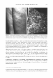
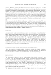
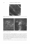
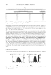
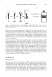
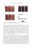
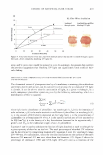
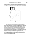
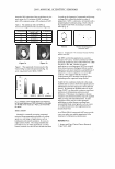
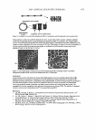
ANALYSIS AND REVIEW OF CELLULITE 387 PRIMOS (FRINGE PROJECTION) PRIMOS is a well known application for measuring changes in skin surface texture and wrinkles. The optical device can also complete 3D measurement of parts of the human body. In instances of measuring an area of 300 x 200 mm, the measurement of the 3D profile is performed with an accuracy of 50 µm. This high accuracy enables application in the cosmetic and medical fields, including dermatology. Furthermore, the technique offers rapid measurements, micron resolution in all axes to ensure the precise determi nation of all measured parameters, highly precise matching of skin areas measured before and after treatment, determination of those characteristics essential for the investigation, and description of body parts with only one instrument. PRIMOS should also detect alterations to the subcutis/cutis structure if this affects the surface topography, i.e., PRIMOS measures the surface irrespective of the causes for changes in skin surface topography. PRIMOS could easily lend application for topical cellulite products devel oped for visualizing surface changes of female thigh skin rather than those products attempting to address the fibrosclerosis or adipose defects. IN VIVO (VIDEO) CONFOCAL MICROSCOPY In vivo confocal microscopy (CFM), using a Vivascope 1500, was employed in a recent study (61) in an attempt to identify specific characteristics of subcutaneous cellulite based on published histopathological findings. The main objective was to compare the impact of cellulite adipose protrusion at the papillary dermis, using in vivo confocal microscopy in the subcutaneous adipose tissue in healthy non-diabetic females, with that of healthy, non-diabetic males. Eleven subjects, five males and six females, were re cruited into the study. Height, weight, and body mass index (BMI) were recorded. All females had visual cellulite on the thighs. In vivo confocal imaging was captured on the front, side, and back aspects of the thighs, 15 images captured per site. Initial results found that striae were seen penetrating into papillary dermis, which were not seen in male skin. Surrounding these "epidermal" striae, which were located and observed within the epidermis at the basal-spinous cell layers, were strands of sclerotic collagen fibers (Figure 4). In contrast, collagen observed in males at the same thigh point was normal-sparse and fine with minimal obvious density (Figure 5 ). In males, imaging before loss of microscope resolution extended to 170 microns, whereas in females this was extended to 200 microns. The average epidermal thickness in males was 5 5 microns compared to 51 microns in females. Larger differences were seen in full-thickness measurements (stratum corneum to the sub-papillary layer of the papillary dermis). However, this may be attributed to loss of resolution in male subjects. Clear differences were observed between females with average BMI and females with greater than average BMI. Females with greater than average BMI had thinner full-thickness measurements (156 microns) than average-BM! females (178 microns). Epidermal thick ness in females with greater than average BMI was less (45.5 microns) than in females with average BMI (62 microns). Collagen as a finely woven meshwork of collagen fibers is found at the papillary layer of the dermis, which includes not only the sub-epidermal papillae situated between the rete ridges but also the sub-papillary layer forming a narrow ribbon between the rete ridges and the sub-papillary blood vessels-papillary dermis. The collagen at this site is
Purchased for the exclusive use of nofirst nolast (unknown)
From: SCC Media Library & Resource Center (library.scconline.org)