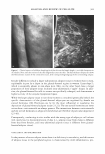
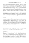
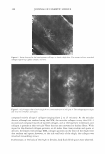
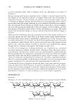
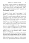
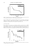
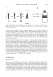
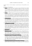
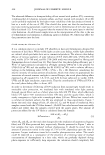
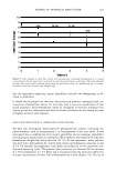
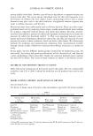
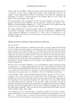
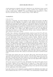
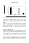
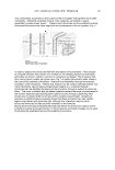
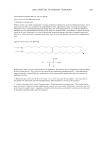
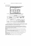
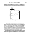
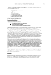
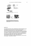
384 JOURNAL OF COSMETIC SCIENCE therefore limited to small scientific studies. However, histopathology is excellent for visualizing cellular, pathological, and biochemical changes in the cellulite condition, and could be recommended as a scientific tool rather than as a commercial tool for cellulite studies. It could also be used as a technique to support image analyses from any of the techniques described herein. ULTRASOUND Much of the clinical aspects of measuring the cellulite condition have relied on echog raphy or ultrasound. Ultrasound measures the acoustic signal recorded from a digital ultrasound wave. Currently, high-resolution ultrasound is a common technique for clinical cellulite imaging. With ultrasound techniques, the skin receives pulses of waves sent through a skin probe. The technique will provide information only on tissue irregularity, rather than on ultrastructural resolution (Figure 2). What is observed is that the dermis is rich in ultrasound reflections (echo-dense), whereas the interphase to the subcutaneous fat, which is echo-poor, is well-defined. This technique also measures the ratio of the subcutis to the surface of the skin because of the protrusion of fat into the dermis. In cellulite the length of the dermal ridge is increased, and comparing this length, which is extended in cellulite to the surface of the epidermis, gives an indication of the severity of adipose tissue protrusion into the dermis. The limitation of the method is that it only indicates a change in adipose tissue thickness rather than categorically demonstrating an actual change in the fibrosclerotic tissue, which would lead to a physical and pathological change in the condition. MAGNETIC RESONANCE IMAGING (MRI) This method has been published in three recent cellulite studies, by Querleux et al. (10) Figure 2. Full-skin-thickness scan (20 MHz) using ultrasound (Cortex Technology ApS, Denmark). The different skin regions are well-defined.

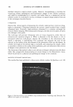
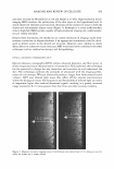
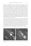
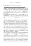
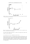
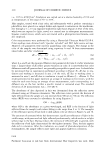
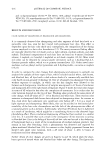
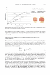
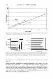
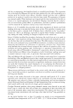
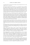
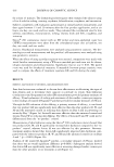
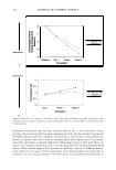
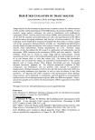
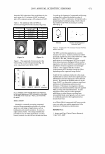
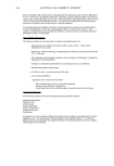
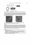
ANALYSIS AND REVIEW OF CELLULITE 385 and more recently by Mirrashed et al. (9) and Smalls et al. (60). High-resolution micro imaging MRI visualizes the architecture of the skin layers at the hypodermal level. It clearly shows the thickness and structural alterations of the connective tissue in both the dermis and subcutaneous adipose tissue (Figure 3). Although it is a very useful method, clinical high-field MRI systems capable of high-resolution imaging are, unfortunately, not yet readily available. Despite these limitations, the method at its current resolution of imaging would dem onstrate a reduction in adipose thickness if an appropriate formulation could be devel oped to deliver actives to the desired site of action. However, since cellulite is clearly about defects in connective tissue structure, MRI would need to be combined with other techniques such as confocal microscopy and histopathology. OPTICAL COHERENCE TOMOGRAPHY (OCT) Optical coherence tomography (OCT) utilizes advanced photonics and fiber optics to obtain images and tissue characterization in human skin. Fully exploited, the technology has the potential to change the way researchers and scientists see and understand the skin. The technology combines the principles of ultrasound with the imaging perfor mance of a microscope. Whereas ultrasound produces images from backscattered sound "echoes," OCT uses infrared light waves that reflect off the internal microstructure within the biological tissues. The frequencies and bandwidths of infrared light are orders of magnitude higher than medical ultrasound signals, resulting in a greatly increased image resolution, 8-25 times greater than that from any other existing modality. Figure 3. Magnetic resonance imaging scans of full-thickness skin (taken from ref. 9). Adipose tissue (b), unlike the dermis (a), is clearly defined.
Purchased for the exclusive use of nofirst nolast (unknown)
From: SCC Media Library & Resource Center (library.scconline.org)