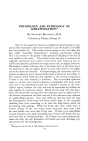
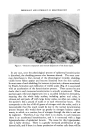
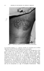
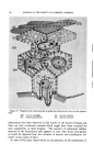
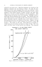
522 JOURNAL OF THE SOCIETY OF COSMETIC CHEMISTS Keratin is the water-insoluble protein present in horny structures. Kera- tinization is the formation of this water-insoluble protein. Epidermal keratinization and differentiation of the horny layer are not synonymous terms, because the horny layer contains not only keratin, but also the degradation products of nucleoproteins, lipoproteins and of other cellular proteins. At least one-fifth of the horny layer consists of substances other than keratin. Therefore chemical analyses of whole epidermal scales do not reflect the process of keratinization alone, but also other processes of cellular differentiation which occur during the formation of the horny layer. It is essential to always keep in mind that "horny layer" and "keratin" are not identical terms. Many errors in interpreting experimental data in pathological conditions are due to the failure to distinguish between these two terms. Profound as the physicochemical changes are during epidermal keratini- zation, the study of their individual stages is very difficult. The main difficulty is that the separation of the individual epidermal components is at best a crude procedure, because of the thinness of the epidermis. To overcome these difficulties, several approaches have been tried: 1. Animal specimens with a thick epidermis were used. Most suc- cessful of these attempts was the study of the epidermis from the cow's nose or lip. From this tissue with 6M urea, Rudall extracted a fibrous protein, epidermin. Epidermin has an x-ray diffraction pattern like keratin and a relatively low sulfur content. It shows increasing consolida- tion as it progresses toward the more superficial layers and is classified in the general group of fibrous proteins to which keratin, myosin and fibrino- gen belong (1). Interesting as these findings are, their significance for the study of human epidermal keratinization has been greatly overrated. Epidermin has never been isolated from human epidermis when extracted with 6M urea, human epidermis yields no specific urea-soluble protein (2). 2. A convenient way would be to rely solely on histological or histo- chemical observations. However, such studies have severe limitations. To cite one common error, one only has to recall the word "keratohyalin." Keratohyalin is the clumped basophilic material in the cytoplasm of the cells of the granular layer, underneath the horny layer. The name im- plies that it is related to keratin. It has been claimed that keratohyalin is a forerunner of keratin, on no particular evidence, except because of its proximity to the horny layer. However, its role in keratinization is most uncertain, because it occurs in nonkeratinizing structures and is absent in some types of keratinization, such as nail formation. Histochemical data must be interpreted with equally great caution. Misinterpretation of specimens stained with nonspecific sulfhydryl stains

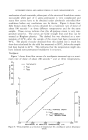
PHYSIOLOGICAl, AND PATHOLOGICAL EPIDERMAI, KERATINIZATI•)N 523 has led to a great deal of confusion in our concepts of epidermal keratini- zation. 3. We are therefore left with straight chemical studies. A simple and ingenious approach has been suggested by Ward and Lundgren in their brilliant review of keratinization (3). These authors attempted to answer the following question: What makes keratins different from other pro- teins ? Ward and Lundgren started with the assumption that epidermal kera- tinization is the most basic type of keratinization. Other types, such as nail or hair formation, are merely modifications of this basic process, by the addition of some components to the basic keratin molecule. Such an addition will lead to harder and more consolidated products. There- fore, if we compare epidermal keratin with hair or nail on the one hand and with a so-called average protein on the other hand, we can set up a series. This series represents something akin to a phylogenetic series the average protein is the most fundamental structure, the soft epidermal keratin the simplest modification of it, while hard keratins represent the most advanced horny structures in this series. By following the trend in amino acid composition from an average protein to a hard keratin, we find that epidermal keratins undergo three types of adaptations: primary and secondary adaptations and those of unknown significance. Primary adaptations are considered to be the essential changes that characterize keratinization as a well-defined process. They include de- creases of methionine and serine and increases in cystine and arginine as we proceed from the softest to the hardest structures. Serine and roethio- nine are involved in the synthesis of cystine and therefore their decrease goes hand in hand with an increase in cystine content. Arginine, with the two other basic amino acids, lysine and histidine, is considered to be part of the basic framework of the keratin molecule. On the basis of the histidine-lysine-arginine ratios, Block divided the keratins into the well- known two groups of eukeratins, with a ratio of 1:4:12 and pseudokeratins with a ratio of 1:3:4. These ratios vary within wide limits and are not as well defined as originally postulated. The secondary adaptations affect the essential amino acids phenyl- alanine, histidine and tryptophane. These amino acids decrease in the course of phylogenesis as the average protein is transformed to keratin. This decrease may be considered a physiologic adaptation to conserve the essential amino acids. By virtue of these adaptations, the keratin which is cast off from the skin surface contains fewer of these essential amino acids. Finally, of unknown significance are decreases in proline and threonine. 4. Interesting as this approach is, it does not shed any light on the process of epidermal keratinization itself. For the study of this process,
Purchased for the exclusive use of nofirst nolast (unknown)
From: SCC Media Library & Resource Center (library.scconline.org)