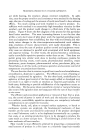
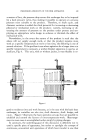
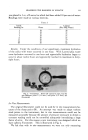
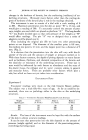
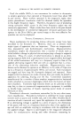
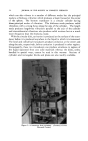
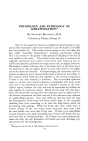
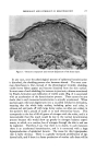
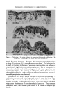
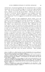
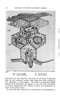
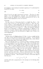
590 JOURNAL OF THE SOCIETY OF COSMETIC CHEMISTS point to a continuing analogy between muscle and epidermis on levels other than that of the polypeptide chain configuration of their fibrous pro- teins. In basal cells some filaments may always be found contiguous with the external nuclear membrane. This is not the rule in the next succeeding layers where there is frequently considerable space between this nuclear membrane and the nearest tonofibril as illustrated in Fig. 1. Mito- chondria are commonly found in this area between the nucleus and tono- filaments in the typical "prickle" cell of the higher Malpighian layers. It is almost impossible to make valid quantitative comparison of ma- terial in ultrathin sections, but study of the electron micrographs does indicate that the actual number of tonofilaments is the same in the basal as in the next succeeding layers. This is particularly clear in Fig 1. This observation coupled with the fact that filaments and the nuclear mem- brane appear contiguous only in the basal layer suggests, admittedly on rather flimsy grounds, that the filaments are synthesized in the basal layer. All other cell organelles are the same in the basal as in the more super- ficial Malpighian cells. These include mitochondria, nuclei, endoplasmic reticulum, cytoplasmic particulates and desmosomes. Submicroscopic cytoplasmic particulates (31) are prominent, reflecting the well-kno•vn basophilia of these cells. The endoplasmic reticulum (32) is, however, very sparse so that the particulates are most commonly distributed freely throughout the cytoplasm without adsorption on membranes or tubules. In footpad skin, the tonofibrillae are exceedingly prominent while in thin skins the filaments may remain widely dispersed without strong organiza- tion into fibrils. This variation obviously reflects the occasions when tonofibrillae can or cannot be detected in these cells by light microscopic techniques. The "prickle" or "spinous" appearance of these cells is due to separation of adjacent cell membranes from each other in the area be- tween desmosomes, i.e., cell membranes are closelyj oined within desmosom es but, due to tension and/or fixation artefacts may separate from each other in the area in between thus giving rise to the scallopped effect. In pig- mented skins, pigment granules may be found within these cells although they are more common within basal cells where, as light microscopic studies have indicated, they frequently localize at one pole of the nucleus. The granules appear noncrystalline (i.e. without sharp, oriented crystal faces) and extremely dense. Occasionally, in particularly thin sections, they appear to be made up of fine granular particulates in an unoriented mass, as noted by others (33) and illustrated in Fig. 2. Stratum granu/osum The transition between the Malpighian and granular laver is marked bv

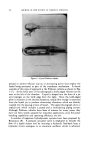
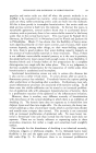
HUMAN EPIDERMIS REVEALED BY ELECTRON MICROSCOPE 591 an increased density and granularity of cytoplasm. In the stratum granulosum large dense irregularly shaped masses whose size ranges from the microscopic to the submicroscopic are present throughout the cyto- plasm. By virtue of their size, density and distribution, these bodies cor- respond to the microscopically prominent "keratohyalin" granules. They are completely nonspecific in size and contour and display no regular internal structure, so that their morphology is consistent with the wide- spread belief that they represent a particular type of cellular debris or side- product in keratinization. In the thin skins (abdomen, breast and fore- skin) that were examined in this study, cells of the Malpighian type develop directly into cornified cells apparently identical to that of the plantar skin type. However, submicroscopic granules, similar to those identified as keratohyalin granules in the plantar skin, have been observed in the cell layer immediately below the cornified one in these thin skins as well. This observation would suggest that keratohyalin granules may be a normal feature of all epidermal types, being small and hence invisible to light microscopy in thin skins and where keratinization is slow. They may remain small in the latter cell types because in these cases there is more time and opportunity for the cell to get rid of its by-products. It is difficult to study these granules in the heavily fibrous epidermis. More information might be gained concerning them in mucous membranes or in the skins of other species. Filaments, closely packed into fibrils, are the only other conspicuous cytoplasmic component of cells of the true S. granulosum. The absence of recognizable mitochondria or submicroscopic (RNA) particulates is rendered less significant by the fact that fixation must be poor in these highly osmiophilic and fibrous cells which are wedged between the rela- tively impermeable S. corneum and many Malpighian cell layers. How- ever, in view of the fact that the nuclei appear well preserved and there is universal agreement that RNA basophilia is markedly decreased (34) and other cytoplasmic components are almost completely degenerated or absent in these cells, this electron microscopic evidence of their absence, can be accepted. It is in this cell layer that the filaments themselves undergo a change. Most fibrils are of the same type as in the lower cell layers with the fila- ments perhaps even more closely packed so that the fibrils appear denser. Some fibrils, however, appear different than the others in being less elec- tron-dense and not possessing filamentous substructure. The two types of fibrils are shown in Fig. 4. In some cases, a fibril of one type appears continuous with that of the other. The conclusion cannot be avoided therefore, that the new hyaline type of fibril is a result of the transforma- tion of the other (the tonofibril) which is taking place in this cell. Desmosomes are particularly prominent in cells of the S. granulosum
Purchased for the exclusive use of nofirst nolast (unknown)
From: SCC Media Library & Resource Center (library.scconline.org)