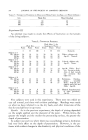
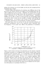
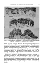
MEASURING THE HARDNESS OF KERATIN 53l than skin softeners per se. As a matter of fact, standard ingredients in many emollient preparations such as hand lotions containing glycerin have perhaps to be discarded if we were to use them primarily as keratin softeners. As is often the case in the introduction of a new concept, the methods used to demonstrate it can be refined and made more exact. This would be a logical step after a principal had been accepted and we attempted to study differences in various chemicals in their ability to make water avail- able to keratin. The purpose of this presentation is primarily to describe and illustrate the two instruments which we used for our in vivo and in vitro studies. The experiments cited merely serve as illustrations for their use. In this paper no attempt has been made to conapare fully the relative merits of the many lanolin fractions or their analogs as water carriers. MATERIALS AND METHODS A practical approach to the problem would be the use of an instrument or instruments of simple construction which were readily available. The senior author in experiments over the last five years has developed an i, vivo and an in vitro method of measuring the hardness of keratin which we believe is a step in the right direction. Very early in our work it became evident that in vitro studies were best carried out by using keratin slices of uniform thickness since we were in- terested in determining how differences in thickness, or the presence or absence of epidermal tissue other than keratin, would effect readings with our instruments. The keratin was obtained from fresh surgical material such as amputa- tions or following autopsy no later than eight to ten hours after death. The tissue was removed with a Brown Dermatome used in skin grafting. This instrument can readily be adjusted so t•'}t an exact thickness of skin can be removed. Tissue was taken from the heel, the arch, and the ball of the foot. Thicknesses used in the experiment were 2ø/1000, 25/1000, aø/1000, 4ø/1000 and 5•/1000 of an inch. Before any experiments were carried out a histological examination was made of the tissue removed. Most of our experiments were carried out with thicknesses of approximately 25/1000 of an inch. Such specimens from our material consisted almost entirely of keratin with but a few scattered islands of epidermal tissue present, if at all. Obviouslyi with a very thi'Ck keratin of the heel at this thickness, we would be dealing with nothing but keratin. INSTRUMENTATION oen Fitro Measurements Blank determined the hardness of his callus by testing its flexibility after

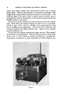
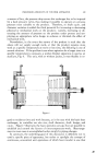
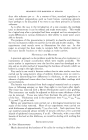
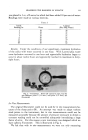
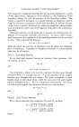
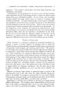
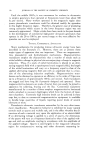
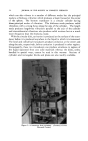
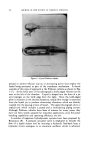
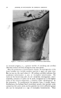
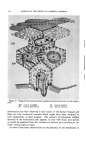
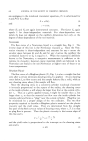
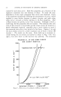
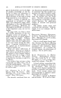
532 JOURNAL OF THE SOCIETY OF COSMETIC CHEMISTS dehydration. While this served the purpose of his experiments, it is ob- vious that flexibility is not always synonymous with hardness. A long enough piece of quite hard material which is not too thick may be flexible and yet quite hard. When a long thin piece of keratin (callus) is made hard by dehydration it will, in nearly all instances, break because of its brittleness when too much bending is attempted. In our experiments we measured the hardness of keratin in a different fashion. The instrument used was a Durometer Hardness Type "A" manufac- tured by the Shaw Instrument Manufacturing Company. It was used in the rubber industry to test the liveliness of rubber materials. As can be seen from the illustration, it is calibrated on a scale of 0 to 100. The re- sistance to penetration by the pin (B) into material depends on the pressure applied to the knob (A). The figure on the scale indicates the depth of penetration and is used for our purpose as an index of hardness (Fig. 1). Figure 1.--Durometer. One of the factors of error is the effect of the resiliency of the underlying surface on the readings. In order to eliminate this, the keratin was placed on a piece of plate glass 0.5 cm. thick. Any cushioning effect of the ma- terial under the keratin was thus eliminated. Maximum pressure on knob (A) on the glass itself gave a reading of 80 on the scale. This was used as a control. Preparation of Material• As soon as the tissue was removed with the Dermatome it was placed between two glass slides to prevent curling at the edges. It was then desiccated by placing in a dehydrater over concentrated sulfuric acid (specific gravity, 1.835). The tissue was left in the dehydrater for seventy- two hours.
Purchased for the exclusive use of nofirst nolast (unknown)
From: SCC Media Library & Resource Center (library.scconline.org)