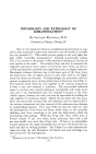
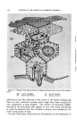
THE FINE STRUCTURE OF HUMAN EPIDERMIS AS REVEALED BY THE ELECTRON MICROSCOPE* t By CEC•I•¾ C^NN^N Sv.I•B¾, PH.D. Department of .4natomy, Cornell Medical College, New York City THE APPLICATION of electron microscopy to ultrathin sections of the epidermis has made it possible to visualize the cell components which are most directly involved in keratinization and desquamation. Since these components, although larger than molecules, are too small to be resolved by light microscopy, information about them helps bridge the gap between chemical and histological studies of the epidermis. The sub- microscopic granules, filaments and membranes to be described here have sometimes been observed in light microscopic studies either because the submicroscopic units naturally group together into microscopically visible units, or because their dimensions are increased by the precipitation upon them of extraneous material as a result of the action of coarse fixatives. This report will attempt to show, however, that in most cases resolution of their ultrastructure is essential for the understanding of the intrinsic function and composition of these cell components. In the following presentation it will be convenient to describe epidermal ultrastructure by starting at the dermoepidermal junction and progressing out from there to the stratum corneum. To eliminate large numbers of illustrations, most observations have been indicated on Fig. 11. BASEMENT MEMBRA•rV. The basement membrane, as described by light microscopy, actually occupies an area of about one-half micron below the surface of the epi- dermis. This junctional area is shown, in electron micrographs, to con- sist of a rather broad membrane (ca/led the derreal membrane by this author (1) closely applied to the basal surface of the epidermal cell on one side and a layer of narrow (300-400 ]k. wide) fibrils on the other (derreal) side. The epidermal cell cytoplasm is thus separated from the dermis by * Presented at the October 4, 1956, Seminar, New York City. t Work supported in part by National Institutes of Health, National Cancer Institute, U.S.P.H.S. Grant C678-C6, while the author was a member of the staff of the Sloan-Kettering Institute for Cancer Research. 584

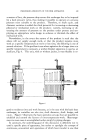
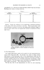
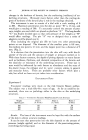
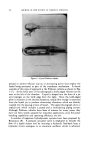
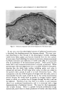
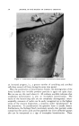
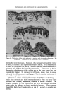
HUMAN EPIDERMIS REVEALED BY ELECTRON MICROSCOPE 585 its own plasma membrane as well as the dermal membrane. There is a space between the two membranes, however, which remains constant at about 350 •. The derreal membrane is about 300 wide and thus about three times as thick as the cell membrane. Electron microscope studies of the basement membrane in amphibian skin (2, 3, 4) and in other tissues have revealed it to consist of two similar components in some cases (7, 8) and of the membranous component only in the kidney (5, 6). In other tissues the membranous component may be considerably thicker than in the epidermis, so that it has been termed the "amorphous" component (8) since, even when quite broad, no internal structure has been seen within it. Relating histochemical studies to these results it becomes clear that the amorphous component (the "dermal membrane" in the epidermis) contains the PAS-positive (mucopolysaccharide) material detectable in this area. Just as the amount of this material is known to vary with histo- logical site (9), the electron microscope shows the thickness of the mem- brane varies, being relatively narrow in the epidermis. Considerable chemi- cal and x-ray diffraction data have accumulated to show that "reticular fibers," such as those at the dermoepidermal junction are actually narrow collagen fibers (10, 11). Thus it comes as no surprise that the filaments adjacent to the dermal membrane display a collagen-like striation in electron micrographs, but are considerably narrower than those in the deeper portions of the dermis. An epidermal cell membrane is always present limiting the epidermal cytoplasm from the basement membrane, and continuous with or con- tiguous to (it has been impossible to distinguish between these two possi- bilities) groups of small dense granules. In tangential sections through the dermal membrane these granules are seen to be distributed in evenly spaced groups. In perpendicular section through the membrane they appear as two rows of 3, 4 or 5 dense spheres to which cytoplasmic fila- ments are attached. At medium magnifications the space between spheres is not resolved, so that the image of a single plate or "bobbin" of electron density is apparent (2, 3, 4). Analysis of a number of angles of section and a number of specimens indicated, however, that these areas actually consist of groups of 3 or 4 short rods or spheres. Cyto- plasmic filaments clearly lead to these bodies and thus terminate there since they never cross over the space between epidermal cell membrane and dermal membrane. This situation is illustrated in the accompanying diagram and was described in detail in an earlier paper (1). Cytoplasmic filaments are generally distributed quite loosely through the cytoplasm of basal cells in thin epidermis while in thick epidermis they are more plentiful and more generally organized into fibrils (compare Figs. 1, 2 and 3). These fibrils are broad enough to be resolved with the light microscope and thus correspond to the tonofibrils which Ranvier
Purchased for the exclusive use of nofirst nolast (unknown)
From: SCC Media Library & Resource Center (library.scconline.org)