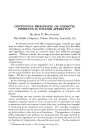
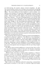
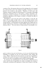
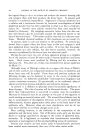
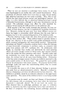
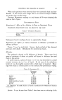
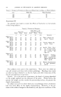
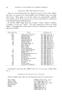
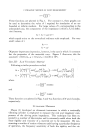
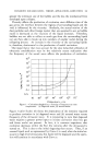
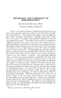
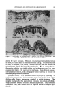
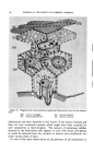
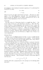
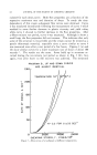
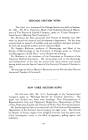
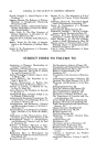
588 JOURNAL OF THE SOCIETY OF COSMETIC CHEMISTS 4-- '•?• ..... ß • .... - ........ --- ½ •-' - 5- 2'•'. --::.•-½. - ':-:.:•::'*' :--• -L: ..&...'.... :' .:.:. :.... :• v •.' '. ................. : .......... ' % .' ....... , ' •,.:• •. v • :.:-. •. ,{:.. %.:.,, • ... • ¾".• ........ . • .,•.,:•, ['% ..... •.. . •½-: ? •:..,. ....... . •,: ,•: ,•- % ...... .t•.. • ... :: ...-- •.:.:% -• . .•' • • ' .,: : .. : '•..b•:" O ': ':1' .: ' '• • ..• • .* " •:' ' '.{: : . - . -- • --' . : -•n ,--• ::-, - -, ,: • •:--- . .... •,:, ½. - •- •- .?:-.- A .- t -• - . :'½ - "-' • . ß . • • % ..: -• .•%-, .•' ',.-,•--• :. •": '.. •' '4 - •x '-• ..-,.....• .:: ,:k. - . •. --%. .: • . - ..... -• .. ..q . - 4::. • ...... ' • '3 ... •h .•.- '::" • • ':.• •' .. • '" ' :. ' • .'•'• ß ( -:•--:,: .*,-.•...:. ...• : ?:-• •...• • :.•-' .• .' • ,., .. • ... [ •-• . - .% .•.' ,. . ,:.-: •-.•.... .:.:- .-- ..."• :'' 2, -:[.• -::: . .,., •' :¾ :: •. ,.• •:. :4 ½-.. .:•-.:::: - .,,,. :.•.• -.. :.. •"• • ..•, .,¾,:•. ' ..... ,•:• ': • ' '7. '•'.. •.-•'• • '"' .,..• •"•' - •' ' 7. ß .• ): ' -.". ,.'.'. •.. : '•. % . ":. ' Plate II, The Transformation of Tonofilaments into Keratin Figure •.--Portion of the cytoplasm of Malpig•an cell in abdominal human skin. Tono- filaments are indicated by arrow. f portion of the nucleus (n) appea• in the lower left-hand

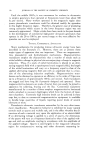
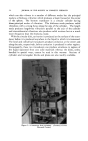
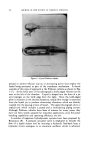
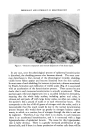
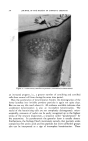
HUMAN EPIDERMIS REVEALED BY ELECTRON MICROSCOPE 589 terminal bars of intestinal epithelia (8) the intercalated discs of cardiac muscle (14) and the intercellular bridges of cervical and vaginal epithelia (12). They are thus present where there is a particular need for cell co 7 hesion, whether or not the cells possess fibrils. Albertini (15) also notes the lack of correlation between desmosomes and tonofibrils since des- mosomes may occur without tonofibrils in anaplastic epidermis. The term "desmosome" (8, 14) is suggested to describe this structure wherever it is found. Before the advent of high magnification optical studies, x-ray dif- fraction and chemical studies had indicated the presence of a fibrous "al- pha" type protein in Malpighian layer cells (16, 17, 18). In addition, a fibrous protein was isolated from a particular type of epidermis (19) with the "alpha" type characteristics of keratin (20). Although this par- ticular protein cannot be extracted from human epidermis it is clear that some alpha-type fibrous protein can be extracted from this tissue and work is under way to characterize this protein further and improve methods for its extraction (21). Despite this evidence and the suggestion, as early as 1936 (16) that tonofibrils are the site of the "alpha" protein and thus are keratin or the keratin precursor, some investigators have continued to consider tonofibrils as some kind of optical or fixation artefact (22, 23). Electron microscope observations indicate that tonofibrils consist of fine o filaments with diameters between 50 and 100 A. (1). Their diameter is thus similar to that of the filaments of one form of keratin (24). From the x-ray diffraction evidence we know that this fibrous protein has a poly- peptide chain configuration similar to that of the other proteins (hair kera- tin, stratum comeurn, fibrinogen, myosin and paramyosin) which also give the "alpha" type wide-angle diffraction diagram (25). Their dimensions are of the same order of magnitude as the filaments in smooth muscle (14, 29) or in the I band of striated muscle (30). It is also interesting to note that both epidermal and cardiac muscle filaments (14) terminate at desmosomes when they reach the plasma membrane. These parallels corner. In addition to a pigment granule (p) and a mitochondrion (m) some endoplasmic reticulum may be seen in the top of the photograph while submicroscopic cytoplasmic particu- lates are distributed throughout the cytoplasm. 26,000 X. Figure &--Comparable area of the cytoplasm of a Malpighian cell in human footpad epi- dermis for comparison with Fig. 2. More tonofilaments are evident (arrow) and these are predominently grouped into tonofibrils. A mitochondrion (m) and submicroscopic particles are also evident. 26,000 X. Figure 4.--Comparable area of the cytoplasm of a cell in the stratum granulosum of the same section from which the photographs of Figs. 3 and 5 were taken. The only cytoplasmic constituents observed are the keratohyalin granules (k) and tonofilaments. The latter are grouped into fibrils, some of which (arrow) have lost electron density and filamentous sub- structure. 26,000 X. Figure 5.--Comparable area of the stratum lucidum adjacent to the cell of Fig. 4. Only .constituent is strands of the nonfilamentous type of fibril (see arrow) which first appeared •n the stratum granulosum (Fig. 4). 26,000 X.
Purchased for the exclusive use of nofirst nolast (unknown)
From: SCC Media Library & Resource Center (library.scconline.org)