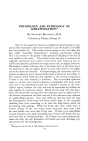
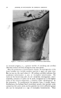
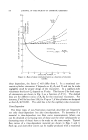
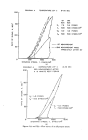
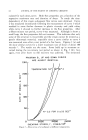
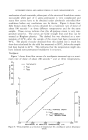
528 JOURNAL OF THE SOCIETY OF COSMETIC CHEMISTS When we turn our attention to pathologic horny layers, we are even more handicapped in our studies than with normal keratinization. First, practically all chemical studies have to be done on scales, because patho- logic epidermal specimens are very rarely available. Then, often no dis- tinction has been made between normal and pathological material. Fi- nally, it is often believed that an abnormal looking horny layer is proof of disturbed keratinization, although in such cases keratinization may be normal. The pathologic process may affect the other metabolic by- products formed during the differentiation of the epidermal cells. Until recently it has been tacitly assumed that psoriasis, the most im- portant of these diseases, was due to qualitatively anomalous keratiniza- tion. However, during the past year, from three different sources evi- dence has begun to accumulate which indicates that the main fault in psoriasis may be in the nonkeratinizing portion of the epidermal cells. The first chemical study of thin skin slices from psoriatic patients was done by Swiss authors (20). These authors found in psoriatic lesions a 50 per cent reduction of the so-called water-soluble polypeptide fraction which contained polypeptides and water-soluble proteins. Subsequently Grfineberg and Szakall in exhaustive analyses found a three-fold decrease of water-extractable components in psoriatic scales, as compared with scales from patients with U.V. erythema (21). In collaboration with Esoda, we extended these studies and determined the water-uptaking ability and amino nitrogen content in whole scales, their aqueous extracts and in the residue left after extraction with water. Psoriatic scales took up considerably less water than callus however, after extraction with water, the residues had all the same water-binding ability. Diminution of the free amino nitrogen content of scales was similarly restricted to the water-soluble portion of the horny layer. We isolated the water-soluble fraction by precipitation with excess acetone. The fraction derived from pathologic scales had much less water-binding ability than its normal counterpart (11, 22). It is possible that most or all of these abnormal findings in psoriatic skin result from an altered proteolytic activity. Recently a marked in- hibition of dipeptidase activity in the affected skin has been observed. The inhibition is probably caused by an unknown substance in the upper layers of the epidermis. The nature of the inhibitory factor is now being studied (23). The question must be raised: are there real diseases of keratinization? Are there diseases with an anomalous horny layer where the keratinous framework is chemically altered? We do not know the answer yet. Most chemical data on pathologic specimens are open to criticism either no distinction has been made between horny layer and keratin, or ' •h"e ' basis of comparison was misleading, because pathologic specimens

PHYSIOLOGICAL AND PATHOLOGICAL EPIDERMAL KERATINIZATION 529 were compared with other pathologic specimens. Often the author starts with a preconceived idea and sets out to prove it. I hope that it has become apparent from my discussion that we cannot afford any preconceived ideas. What we need are facts, data and tech- niques to cope with the many unsolved problems in the field. The findings of the past few years have been most encouraging. There is every reason to believe that within the next few years we will witness the solution of many of our present problems. Progress in this most important area of skin physiology will ultimately benefit the fields of dermatology'. and cosmetic chemistry alike by laying a solid foundation for future treatment techniques. SUMMARY Epidermal keratinization is one of many processes occurring during the development of the horny layer. Because of the thinness of the epi- dermis, satisfactory separation of the individual components during the various stages of this process is not possible. The following approaches have been tried: 1. Chemical studies of animal specimens with a thick epidermis (cow's nose) are not directly applicable to human epidermis. 2. Histological and histochemical studies have inherent limitations and errors. 3. Comparison of epidermal keratins with hard keratins (hair, nails) and with average body proteins have given an insight into the changes during the phylogenetic development of the keratins. 4. Comparison of the amino acid composition of whole human epi- dermis and of the horny layer revealed that during keratinization the over- all chemical change is slight. The only definitely established differences are the disappearance of hydroxyproline and an inconstant and variable increase in cystine during keratinization. At present the best working theory to account for the changes during keratinization is as follows: keratinization is a two-step process. The first step is the formation of a fibrous precursor in the cellular layer of the epidermis, with little or no sulfur. This precursor then combines with sulfur containing amino acids, polypeptides or proteins to form the con- solidated keratin. Studies of pathologic keratinization are often based on the erroneous assumption that an anomalous horny layer necessarily reflects a faulty process of keratinization. About one-fifth of the horny layer consists of nonkeratinous, water-soluble substances, especially free amino acids. Many of these components have a strong water-binding ability. The reduced amino nitrogen and water-binding ability of some pathologic scales is due to alterations in the nonkeratinous portion of the horny scales.
Purchased for the exclusive use of nofirst nolast (unknown)
From: SCC Media Library & Resource Center (library.scconline.org)