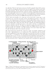
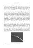
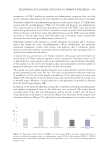
449 DELIVERING SUSTAINABLE SOLUTIONS TO IMPROVE WELLBEING
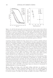
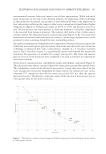
for the reference compound. In general, degradation is followed by determining parameters
like DOC, CO
2 production, and oxygen uptake. The pass levels for ready biodegradability
are 70% removal of DOC and 60% of theoretical oxygen demand or ThCO
2 production for
respirometric methods. These pass values must be reached in a 10-day window within the
28-day period of the test.1
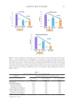
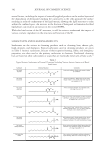
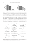
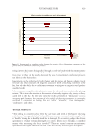
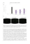
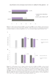
INHIBITION OF HIGH MOBILITY GROUP PROTEIN B1 (HMGB1) STRESSORIN RELEASE IN HUMAN
EPIDERMAL KERATINOCYTES
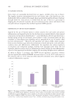
Human epidermal keratinocytes from adults (HEKa cells) were seeded in 96-well plates in
culture medium. After 24 hours of incubation, the culture medium was removed, and a
metabolic stress was applied by adding 20 mM of glucose (Merck Life Science) and 0.1 mM
of palmitic acid (Merck Life Science) to the culture medium, simulating an unhealthy diet
in cell culture with or without treatment with a THW biotech ingredient concentrate
(without glycerin) at 100 µg/mL. Cells treated with culture medium, glucose, and palmitic
acid were used as a metabolic stress control. Cells treated with culture medium alone were
used as a control. After 24 hours of incubation, the supernatants were collected, and the
stressorin inhibition was measured using a stressorin marker (HMGB1) by ELISA Novus
Biologicals, Abingdon, United Kingdom) according to the manufacturer’s protocol. These
results were normalized with the total viable cell number for each condition calculated
by the PrestoBlueTM staining assay (Thermo Fisher Scientific, Waltham, MA, USA). The
obtained values were normalized compared to the control or metabolic stress control
as applicable and represent the mean of three independent experiments performed in
triplicates. The statistical analysis used was the unpaired student’s t test.
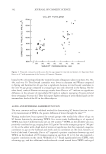
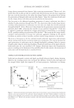
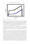
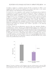
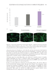
EVALUATION OF THE ANTIGLYCATION EFFECT
To assess the ability of the active ingredient to counteract the formation of advanced glycation
end products (AGEs), an in tubo glycation reaction was performed. Type I collagen (0.6 mg/
mL) was incubated with glucose at 0.4 M and with 100 µg/mL of THW biotech ingredient
concentrate at 60°C for 3 days. As a glycation control, only type I collagen and glucose at
the same concentrations were incubated, a condition in which glycation occurs. Glycation
levels were determined by measuring AGE-specific fluorescence, or 370(ex)/440(em) using
ClarioSTAR. Samples were tested in triplicates and assayed in four different replicates.
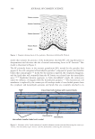
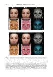
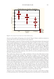
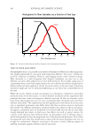
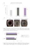
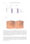
REDUCTION OF LIPOFUSCIN ACCUMULATION WITH AGE IN HUMAN EPIDERMAL KERATINOCYTES
Cellular garbage reduction was evaluated by analyzing lipofuscin accumulation (an
autofluorescent pigment commonly used as an indicator of waste accumulation) in
epidermal cells using autofluorescence. HEKa cells were seeded in 96-well cell carrier
black plates (PerkinElmer, Inc., Waltham, MA, USA) in culture medium. After 24 hours
of incubation, the culture medium was removed, and cells were incubated in culture
medium in combination with metabolic stress (20 mM glucose and 0.1 mM palmitic
acid), simulating an unhealthy diet in the cell culture with or without treatment with
the THW biotech ingredient concentrate at 100 µg/mL. At the same time, cells treated
with the culture medium alone were used as a control, and cells treated with culture
for the reference compound. In general, degradation is followed by determining parameters
like DOC, CO
2 production, and oxygen uptake. The pass levels for ready biodegradability
are 70% removal of DOC and 60% of theoretical oxygen demand or ThCO
2 production for
respirometric methods. These pass values must be reached in a 10-day window within the
28-day period of the test.1
INHIBITION OF HIGH MOBILITY GROUP PROTEIN B1 (HMGB1) STRESSORIN RELEASE IN HUMAN
EPIDERMAL KERATINOCYTES
Human epidermal keratinocytes from adults (HEKa cells) were seeded in 96-well plates in
culture medium. After 24 hours of incubation, the culture medium was removed, and a
metabolic stress was applied by adding 20 mM of glucose (Merck Life Science) and 0.1 mM
of palmitic acid (Merck Life Science) to the culture medium, simulating an unhealthy diet
in cell culture with or without treatment with a THW biotech ingredient concentrate
(without glycerin) at 100 µg/mL. Cells treated with culture medium, glucose, and palmitic
acid were used as a metabolic stress control. Cells treated with culture medium alone were
used as a control. After 24 hours of incubation, the supernatants were collected, and the
stressorin inhibition was measured using a stressorin marker (HMGB1) by ELISA Novus
Biologicals, Abingdon, United Kingdom) according to the manufacturer’s protocol. These
results were normalized with the total viable cell number for each condition calculated
by the PrestoBlueTM staining assay (Thermo Fisher Scientific, Waltham, MA, USA). The
obtained values were normalized compared to the control or metabolic stress control
as applicable and represent the mean of three independent experiments performed in
triplicates. The statistical analysis used was the unpaired student’s t test.
EVALUATION OF THE ANTIGLYCATION EFFECT
To assess the ability of the active ingredient to counteract the formation of advanced glycation
end products (AGEs), an in tubo glycation reaction was performed. Type I collagen (0.6 mg/
mL) was incubated with glucose at 0.4 M and with 100 µg/mL of THW biotech ingredient
concentrate at 60°C for 3 days. As a glycation control, only type I collagen and glucose at
the same concentrations were incubated, a condition in which glycation occurs. Glycation
levels were determined by measuring AGE-specific fluorescence, or 370(ex)/440(em) using
ClarioSTAR. Samples were tested in triplicates and assayed in four different replicates.
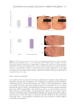
REDUCTION OF LIPOFUSCIN ACCUMULATION WITH AGE IN HUMAN EPIDERMAL KERATINOCYTES
Cellular garbage reduction was evaluated by analyzing lipofuscin accumulation (an
autofluorescent pigment commonly used as an indicator of waste accumulation) in
epidermal cells using autofluorescence. HEKa cells were seeded in 96-well cell carrier
black plates (PerkinElmer, Inc., Waltham, MA, USA) in culture medium. After 24 hours
of incubation, the culture medium was removed, and cells were incubated in culture
medium in combination with metabolic stress (20 mM glucose and 0.1 mM palmitic
acid), simulating an unhealthy diet in the cell culture with or without treatment with
the THW biotech ingredient concentrate at 100 µg/mL. At the same time, cells treated
with the culture medium alone were used as a control, and cells treated with culture