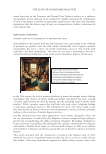
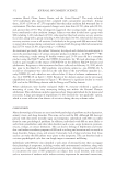
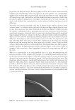
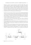
465 DELIVERING SUSTAINABLE SOLUTIONS TO IMPROVE WELLBEING
enhanced production of hyaluronic acid in the skin and not only increases skin hydration
but also promotes many healing aspects and tissue remodeling. S rebaudiana extract was
also shown to stimulate skin renewal and repair as it boosted HB-EGF, a member of the
family of epidermal growth factors required for re-epithelialization processes. These results
suggest that S rebaudiana extract is involved in skin regeneration.
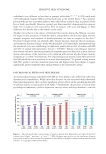
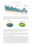
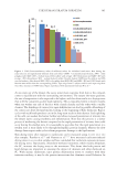
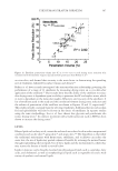
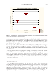
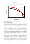
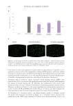
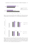
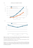
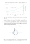
To further prove the retinoid-like efficacy, the ability of S rebaudiana extract to regulate
dermis remodeling was tested. S rebaudiana extract behaved similarly to retinoic acid
(Figure 14) as the collagen and elastin levels were boosted and MMPs were reduced, which
helps preserve the dermis for a rejuvenating effect.
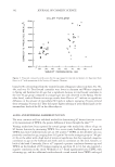
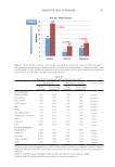
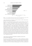
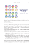
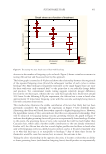
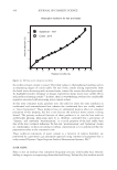
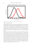
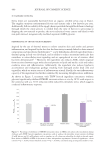
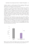
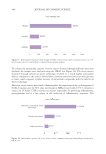
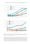
Finally, to provide additional confirmation of retinoid-like efficacy, the extract’s ability to
boost the antioxidant response of skin cells was tested (Figure 15). S rebaudiana extract
increased the expression of HMOX1 by 954%, the expression of TXNRD1 by 209%,
and the expression of oxidative stress induced growth inhibitor 1 (OSGIN1) by 261%,
respectively. These genes are involved in cellular protection against oxidative stress,
detoxification of reactive oxygen species, and redox signaling.
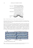
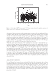
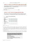
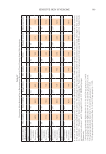
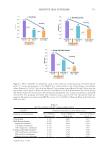
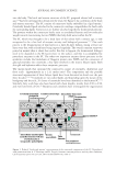
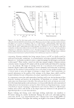
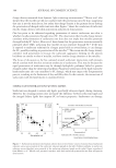
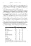
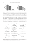
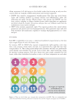
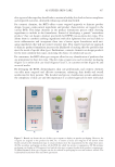
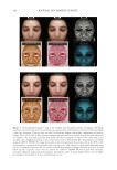
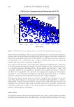
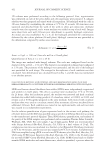
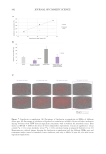
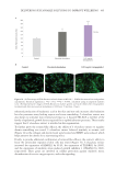
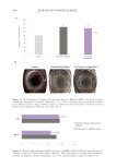
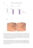
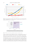
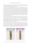
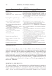
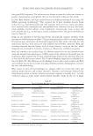
Figure 10. (A) Percentage of EDA-fibronectin levels shown as MEAN ± SEM of the mean of four independent
experiments. Statistical significance: **p 0.01, ****p 0.0001, calculated using an unpaired student’s
t-test. (B) Representative images of EDA-fibronectin marker (green) and nuclei (blue) after tetrapeptide-1
treatment and after microcurrent stimulation (1000 µA, 70 mv/mm, 1 hour).
enhanced production of hyaluronic acid in the skin and not only increases skin hydration
but also promotes many healing aspects and tissue remodeling. S rebaudiana extract was
also shown to stimulate skin renewal and repair as it boosted HB-EGF, a member of the
family of epidermal growth factors required for re-epithelialization processes. These results
suggest that S rebaudiana extract is involved in skin regeneration.
To further prove the retinoid-like efficacy, the ability of S rebaudiana extract to regulate
dermis remodeling was tested. S rebaudiana extract behaved similarly to retinoic acid
(Figure 14) as the collagen and elastin levels were boosted and MMPs were reduced, which
helps preserve the dermis for a rejuvenating effect.
Finally, to provide additional confirmation of retinoid-like efficacy, the extract’s ability to
boost the antioxidant response of skin cells was tested (Figure 15). S rebaudiana extract
increased the expression of HMOX1 by 954%, the expression of TXNRD1 by 209%,
and the expression of oxidative stress induced growth inhibitor 1 (OSGIN1) by 261%,
respectively. These genes are involved in cellular protection against oxidative stress,
detoxification of reactive oxygen species, and redox signaling.
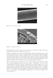
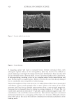
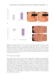
Figure 10. (A) Percentage of EDA-fibronectin levels shown as MEAN ± SEM of the mean of four independent
experiments. Statistical significance: **p 0.01, ****p 0.0001, calculated using an unpaired student’s
t-test. (B) Representative images of EDA-fibronectin marker (green) and nuclei (blue) after tetrapeptide-1
treatment and after microcurrent stimulation (1000 µA, 70 mv/mm, 1 hour).