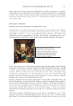
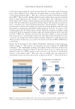
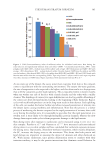
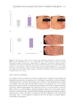
341 Aging Skin Barrier
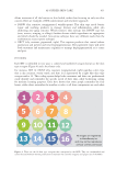
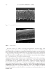
surface.20–23 Corneocytes are flat hexagonal or pentagonal cells about 25 µm on a side, with
a surface area ranging from about 550–1300 µm2 depending on age and body site,24–27
and are about 0.5–1.0 µm thick.26,28 Ceramides and free fatty acids are bound to the outer
surface of the corneocyte cell envelope.29,30
In the SC filaggrin is released from the microfibrils31 and digested by proteolytic enzymes
to produce key amino acid components of SC’s natural moisturizing factor (NMF).32–36
NMF consists of lactate, amino acids from filaggrin breakdown and pyrrolidone carboxylic
acid (PCA) formed from the amino acid glutamine.37–40 NMF is vital for maintenance of
proper hydration of the SC allowing it to be flexible and to desquamate properly.34,40–44
The SC is 12–16 cell layers thick on most body sites, but it can vary from 9 cell layers on
the forehead or eyelids to as much as 25 on the dorsum of the hand and 50 or more on the
palms or the soles.45
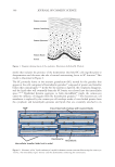
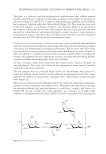
The lipids that are released into the intercellular space as the SC forms are glucosyl ceramides,
cholesterol, cholesterol sulfate, cholesterol esters, and long chain fatty acids. In the intercellular
space the glucosyl ceramides are converted to ceramides.46,47 Ceramides are polar lipids, but
much less hydrophilic than the phospholipids of the original cell membrane. Phospholipids
from the keratinocytes of the viable layers are broken down by phospholipases in the lower
SC to release long chain fatty acids.48–50 Another key transformation in the extracellular
space is the conversion of cholesterol sulfate to cholesterol by steroid sulfatase.51–53 After this
extracellular processing, SC lipids spontaneously organize into multiple layers between the
SC cells.54 This layered structure is critical to the barrier function of the skin.
AGING AND THE STRUCTURE OF THE STRATUM CORNEUM
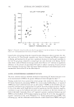
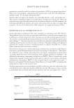
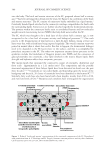
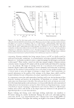
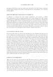
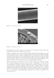
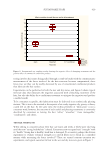
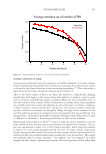
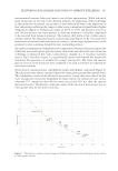
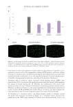
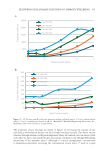
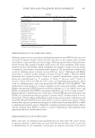
A general trend for corneocytes to increase in surface area with age has been reported by
several groups.25,27,55–57 The reported increase in surface area between young adults and
adults over 55 ranges from 0–14% depending on body site and method of measurement.
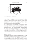
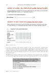
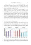
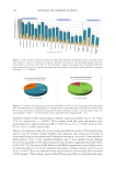
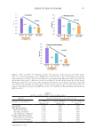
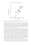
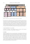
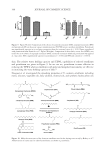
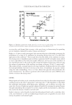
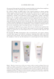
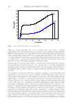
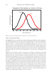
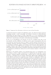
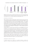
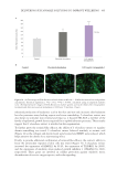
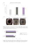
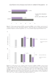
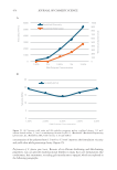
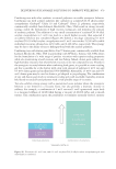
Figure 3 shows corneocyte size data from two different age groups reported by Grove
et al.56 for the volar forearm.
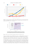
Note the extreme variability in the data from the older age group compared to the narrower
distribution in the younger group. The average size of corneocytes was increased in the
older age group but there is considerable overlap between the two data sets. Starr et al.58
reported a significant increase in corneocyte area with age on the arms of post-menopausal
women compared to premenopausal but no change in corneocyte surface area on the hand.
Imokawa et al. reported a gradual decrease in total SC ceramides with age.59 Rogers et
al. reported progressive reductions in fatty acids, ceramides, and cholesterol with age in
subjects ranging in age from 21–60 years on the hands, legs, and face.60 The reduction in
ceramide I (EOS) linolate was especially noteworthy. Denda et al.61 reported that ceramide
2 decreased and ceramide 3 increased with age after age 40 with female subjects but not
male subjects. Wohlrab et al.,62 investigated changes in free fatty acids between subject
ranging in age from 18–40 to subjects over 60. Only two long chain fatty acids, C15:0 and
C17:0 were found to be reduced in the older age group. Starr et al.58 utilized time-of-flight
secondary ion mass spectrometry (TOF-SIMS) to analyze age-related changes in SC lipids
in female subjects before and after menopause. They reported an increase in cholesterol
sulfate in the older age group in both sun-exposed (hand) and protected (under the arm)
body sites. Fujiwara et al.63 investigated the effect of age and season on ceramide covalently
surface.20–23 Corneocytes are flat hexagonal or pentagonal cells about 25 µm on a side, with
a surface area ranging from about 550–1300 µm2 depending on age and body site,24–27
and are about 0.5–1.0 µm thick.26,28 Ceramides and free fatty acids are bound to the outer
surface of the corneocyte cell envelope.29,30
In the SC filaggrin is released from the microfibrils31 and digested by proteolytic enzymes
to produce key amino acid components of SC’s natural moisturizing factor (NMF).32–36
NMF consists of lactate, amino acids from filaggrin breakdown and pyrrolidone carboxylic
acid (PCA) formed from the amino acid glutamine.37–40 NMF is vital for maintenance of
proper hydration of the SC allowing it to be flexible and to desquamate properly.34,40–44
The SC is 12–16 cell layers thick on most body sites, but it can vary from 9 cell layers on
the forehead or eyelids to as much as 25 on the dorsum of the hand and 50 or more on the
palms or the soles.45
The lipids that are released into the intercellular space as the SC forms are glucosyl ceramides,
cholesterol, cholesterol sulfate, cholesterol esters, and long chain fatty acids. In the intercellular
space the glucosyl ceramides are converted to ceramides.46,47 Ceramides are polar lipids, but
much less hydrophilic than the phospholipids of the original cell membrane. Phospholipids
from the keratinocytes of the viable layers are broken down by phospholipases in the lower
SC to release long chain fatty acids.48–50 Another key transformation in the extracellular
space is the conversion of cholesterol sulfate to cholesterol by steroid sulfatase.51–53 After this
extracellular processing, SC lipids spontaneously organize into multiple layers between the
SC cells.54 This layered structure is critical to the barrier function of the skin.
AGING AND THE STRUCTURE OF THE STRATUM CORNEUM
A general trend for corneocytes to increase in surface area with age has been reported by
several groups.25,27,55–57 The reported increase in surface area between young adults and
adults over 55 ranges from 0–14% depending on body site and method of measurement.
Figure 3 shows corneocyte size data from two different age groups reported by Grove
et al.56 for the volar forearm.
Note the extreme variability in the data from the older age group compared to the narrower
distribution in the younger group. The average size of corneocytes was increased in the
older age group but there is considerable overlap between the two data sets. Starr et al.58
reported a significant increase in corneocyte area with age on the arms of post-menopausal
women compared to premenopausal but no change in corneocyte surface area on the hand.
Imokawa et al. reported a gradual decrease in total SC ceramides with age.59 Rogers et
al. reported progressive reductions in fatty acids, ceramides, and cholesterol with age in
subjects ranging in age from 21–60 years on the hands, legs, and face.60 The reduction in
ceramide I (EOS) linolate was especially noteworthy. Denda et al.61 reported that ceramide
2 decreased and ceramide 3 increased with age after age 40 with female subjects but not
male subjects. Wohlrab et al.,62 investigated changes in free fatty acids between subject
ranging in age from 18–40 to subjects over 60. Only two long chain fatty acids, C15:0 and
C17:0 were found to be reduced in the older age group. Starr et al.58 utilized time-of-flight
secondary ion mass spectrometry (TOF-SIMS) to analyze age-related changes in SC lipids
in female subjects before and after menopause. They reported an increase in cholesterol
sulfate in the older age group in both sun-exposed (hand) and protected (under the arm)
body sites. Fujiwara et al.63 investigated the effect of age and season on ceramide covalently