386 JOURNAL OF COSMETIC SCIENCE
contrast, with nonionic and zwitterionic surfactants which are milder towards proteins,
lipid interactions may play a dominant role.
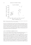
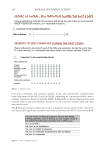
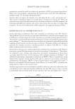
Recently, in vitro systems based on reconstructed skin equivalent models have also become
popular for assessing the skin irritation potential of surfactants and other actives.43–45
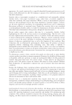
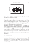
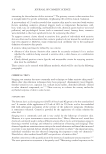
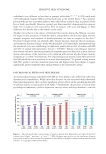
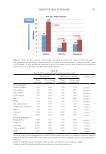
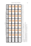
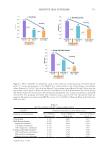
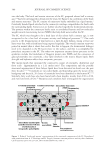
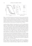
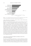
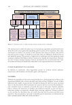
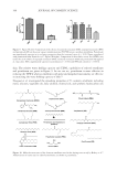
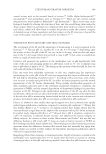
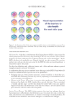
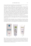
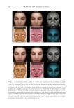
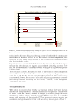
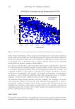
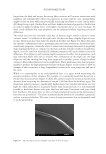
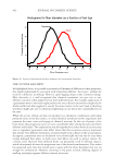
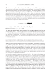
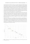
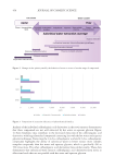
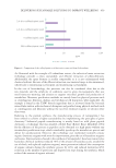
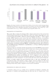
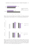
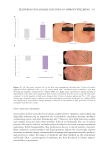
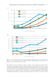
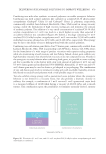
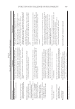
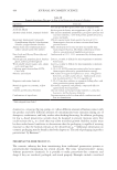
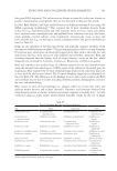
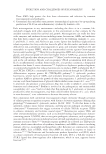
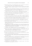
Walters et al. used the 3-D EpiDerm™ model system to evaluate tissue viability and
primary cytokine interleukin-1α release to evaluate the potential dermal irritation of 224
nonionic, amphoteric and/or anionic surfactant-containing formulations, or individual
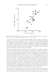
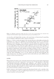
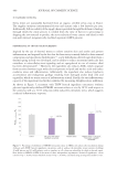
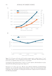
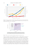
raw materials (see Figure 5).45 The authors showed a correlation between in vivo TEWL
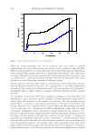
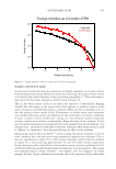
measurements in a patch test and the IL1 alpha release in in-vitro studies (see Figure 6).
The results presented are consistent with the prevailing understanding that the order of
irritation potential follows: anionic amphoteric nonionic. The reasons for differences
within each category were not discussed in this paper. However, the data from such a large
list of surfactants is certainly worth exploring further. Since the quality of the SC barrier in
the EpiDerm model system is relatively weak compared to the real human SC, the results
by such tests systems will be indicative of the inherent irritation potential of an ingredient
rather than in a real-life situation in subjects with healthy SC under normal use conditions.
Since patch tests are rather exaggerated and enhance penetration under an occlusive patch,
the results from such reconstructed models may be reflective of the situation in subjects
with a compromised skin barrier.
STRUCTURE-FUNCTION RELATIONSHIPS GOVERNING SURFACTANT-
INDUCED SKIN IRRITATION
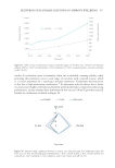
The various test methodologies described above are useful in predicting the irritation
potential of surfactants. It is important at this stage to go beyond just predicting irritation
potential to understanding structure-function relationships governing irritation potential
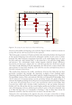
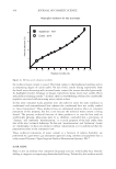
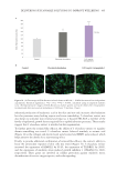
of surfactants. A simplistic analysis by looking at the trends in Figures 3–6 suggests that
the charge and the size of the surfactant head group plays a role in the irritation potential
Figure 5. In vitro assessment of skin irritation potential of surfactant-based formulations by using a 3D skin
reconstructed tissue model and cytokine response. Figure reproduced from Walters et. al.45
contrast, with nonionic and zwitterionic surfactants which are milder towards proteins,
lipid interactions may play a dominant role.
Recently, in vitro systems based on reconstructed skin equivalent models have also become
popular for assessing the skin irritation potential of surfactants and other actives.43–45
Walters et al. used the 3-D EpiDerm™ model system to evaluate tissue viability and
primary cytokine interleukin-1α release to evaluate the potential dermal irritation of 224
nonionic, amphoteric and/or anionic surfactant-containing formulations, or individual
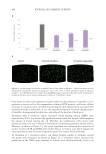
raw materials (see Figure 5).45 The authors showed a correlation between in vivo TEWL
measurements in a patch test and the IL1 alpha release in in-vitro studies (see Figure 6).
The results presented are consistent with the prevailing understanding that the order of
irritation potential follows: anionic amphoteric nonionic. The reasons for differences
within each category were not discussed in this paper. However, the data from such a large
list of surfactants is certainly worth exploring further. Since the quality of the SC barrier in
the EpiDerm model system is relatively weak compared to the real human SC, the results
by such tests systems will be indicative of the inherent irritation potential of an ingredient
rather than in a real-life situation in subjects with healthy SC under normal use conditions.
Since patch tests are rather exaggerated and enhance penetration under an occlusive patch,
the results from such reconstructed models may be reflective of the situation in subjects
with a compromised skin barrier.
STRUCTURE-FUNCTION RELATIONSHIPS GOVERNING SURFACTANT-
INDUCED SKIN IRRITATION
The various test methodologies described above are useful in predicting the irritation
potential of surfactants. It is important at this stage to go beyond just predicting irritation
potential to understanding structure-function relationships governing irritation potential
of surfactants. A simplistic analysis by looking at the trends in Figures 3–6 suggests that
the charge and the size of the surfactant head group plays a role in the irritation potential
Figure 5. In vitro assessment of skin irritation potential of surfactant-based formulations by using a 3D skin
reconstructed tissue model and cytokine response. Figure reproduced from Walters et. al.45