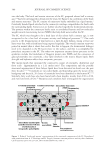
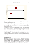
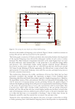
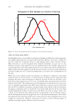
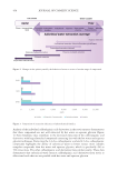
347 Aging Skin Barrier
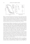
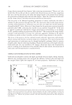
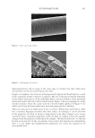
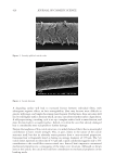
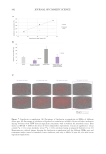
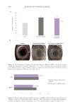
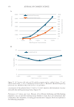
Biniek at al.104 investigated the effect of age on the mechanical properties of isolated SC.
They reported that the SC stiffened with age, and that delamination energy increased with
age. These factors could potentially contribute to an increased tendency to form both cracks
and flakes in elderly skin.
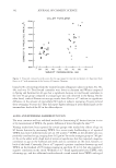
Lactate is a component of the NMF that may be derived either from sweat105 or through
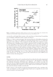
anaerobic metabolism in the upper epidermis.106 Nakagawa et al. investigated the correlation
between NMF components and SC stiffness, pH, and hydration in summer and winter with
healthy subjects.42 The only components that correlated significantly to SC stiffness and
hydration were lactate and potassium. One might expect that the slower metabolism of
aging skin reflected by the lower SC turnover rate might lead to lower levels of lactate in
the SC. However, Prahl and coworkers reported that keratinocytes from aging skin produce
higher levels of lactate ex vivo.107 It is well established that sweating rates decrease with
age108 which might lead to lower levels of lactate and thus stiffer, less well hydrated SC.
Another factor may be the decline sebum production that occurs with age109 especially
in post-menopausal women. Fluhr et al. have presented data from studies of asebic
mice, indicating that glycerol derived from the hydrolysis of sebum contributes to skin
hydration.110 Sebum production declines dramatically in women at menopause but does not
decline significantly in men before age 80109 so this may be relevant to post-menopausal
women but not to men before age 80.
It is also possible that the increased cholesterol sulfate observed in older subjects by Starr
et al.58 contributes to skin scaling. Congenital X-linked ichthyosis results from a defect in
the enzyme that convert cholesterol sulfate to cholesterol51 and there is evidence that this is
the main cause of the excessive skin scaling in this disease52 probably by inhibition of the
enzymes that break down SC desmosomes.111
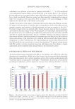
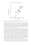
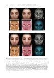
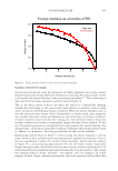
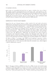
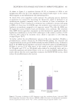
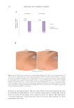
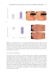
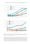
Tagami’s et al. studied the dorsum of the hands of Japanese golfers who only wore a golfing
glove on one hand to compare intrinsic aging to photoaging on properties of the SC.103
Roughness of the skin surface was measured from silicon replicas. Interestingly, there was
a significant negative correlation between the difference in roughness between exposed
and covered hands and the golfer’s handicap. Better golfers (lower handicap) had larger
increases in roughness on their exposed hand. Hydration as measured by higher frequency
conductance was lower for the more exposed site. indicating a drier skin surface but there
was no difference in barrier function as measured by TEWL. Tojahn et al.112 compared sun-
exposed and protected skin on the arms of female subjects and also found no significant
difference between exposed and protected sites.
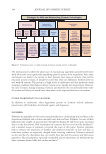
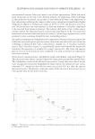
The tendency for elderly subjects to develop dry skin probably has multiple contributing
factors including lower PCA levels, the slower rate of SC renewal, lower levels of lactic acid
due to reduced sweating, possibly reduced glycerol due to reduced sebum production and
perhaps other factors that have yet to be discovered.
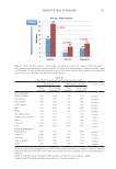
CONCLUSIONS
Aging and photoaging profoundly affect the structure of the dermis leading to well
characterized changes in skin structure and appearance.1–4,113 The effect of age on the
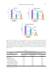
epidermal barrier is less obvious and less pronounced. Somewhat surprisingly there is little
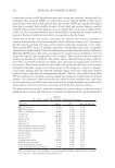
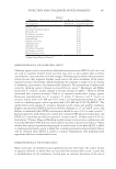
or no decline in SC barrier function as measured by TEWL (see Table I) except for the
décolleté in women68 and one report on the forehead.69 In fact most studies indicate reduced
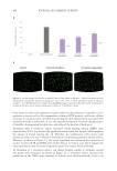
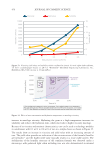
Biniek at al.104 investigated the effect of age on the mechanical properties of isolated SC.
They reported that the SC stiffened with age, and that delamination energy increased with
age. These factors could potentially contribute to an increased tendency to form both cracks
and flakes in elderly skin.
Lactate is a component of the NMF that may be derived either from sweat105 or through
anaerobic metabolism in the upper epidermis.106 Nakagawa et al. investigated the correlation
between NMF components and SC stiffness, pH, and hydration in summer and winter with
healthy subjects.42 The only components that correlated significantly to SC stiffness and
hydration were lactate and potassium. One might expect that the slower metabolism of
aging skin reflected by the lower SC turnover rate might lead to lower levels of lactate in
the SC. However, Prahl and coworkers reported that keratinocytes from aging skin produce
higher levels of lactate ex vivo.107 It is well established that sweating rates decrease with
age108 which might lead to lower levels of lactate and thus stiffer, less well hydrated SC.
Another factor may be the decline sebum production that occurs with age109 especially
in post-menopausal women. Fluhr et al. have presented data from studies of asebic
mice, indicating that glycerol derived from the hydrolysis of sebum contributes to skin
hydration.110 Sebum production declines dramatically in women at menopause but does not
decline significantly in men before age 80109 so this may be relevant to post-menopausal
women but not to men before age 80.
It is also possible that the increased cholesterol sulfate observed in older subjects by Starr
et al.58 contributes to skin scaling. Congenital X-linked ichthyosis results from a defect in
the enzyme that convert cholesterol sulfate to cholesterol51 and there is evidence that this is
the main cause of the excessive skin scaling in this disease52 probably by inhibition of the
enzymes that break down SC desmosomes.111
Tagami’s et al. studied the dorsum of the hands of Japanese golfers who only wore a golfing
glove on one hand to compare intrinsic aging to photoaging on properties of the SC.103
Roughness of the skin surface was measured from silicon replicas. Interestingly, there was
a significant negative correlation between the difference in roughness between exposed
and covered hands and the golfer’s handicap. Better golfers (lower handicap) had larger
increases in roughness on their exposed hand. Hydration as measured by higher frequency
conductance was lower for the more exposed site. indicating a drier skin surface but there
was no difference in barrier function as measured by TEWL. Tojahn et al.112 compared sun-
exposed and protected skin on the arms of female subjects and also found no significant
difference between exposed and protected sites.
The tendency for elderly subjects to develop dry skin probably has multiple contributing
factors including lower PCA levels, the slower rate of SC renewal, lower levels of lactic acid
due to reduced sweating, possibly reduced glycerol due to reduced sebum production and
perhaps other factors that have yet to be discovered.
CONCLUSIONS
Aging and photoaging profoundly affect the structure of the dermis leading to well
characterized changes in skin structure and appearance.1–4,113 The effect of age on the
epidermal barrier is less obvious and less pronounced. Somewhat surprisingly there is little
or no decline in SC barrier function as measured by TEWL (see Table I) except for the
décolleté in women68 and one report on the forehead.69 In fact most studies indicate reduced