340 JOURNAL OF COSMETIC SCIENCE
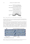
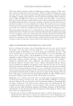
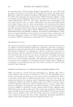
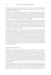
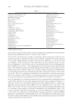
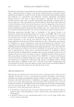
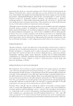
model that includes the presence of the desmosomes that hold SC cells together prior to
desquamation and discusses the role of natural moisturizing factor in SC function.9 This
model is illustrated in Figure 2.
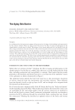
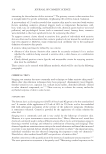
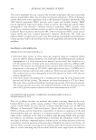
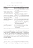
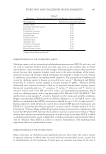
The SC primarily forms in the stratum granulosum (SG), named for the granules that
appear in the cells comprised of keratohyalin granules10 composed of protein and lamellar
bodies that contain lipids.11,12 In the SG: the nucleus is digested, the cytoplasm disappears,
and the lipids that will eventually form the SC barrier are released into the intercellular
space.7,13–15 Epidermal keratins aggregate to form microfibrils16 inside the corneocytes
under the influence of filaggrin from the keratohyalin granules.17–19 The keratinocyte cell
membrane is replaced by the corneocyte cell envelope made of cross-linked protein from
the cytoplasm and keratohyalin granules and lipids that are covalently attached to its
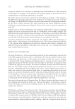
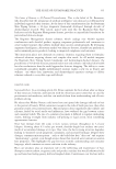
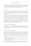
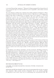
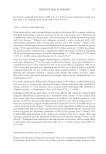
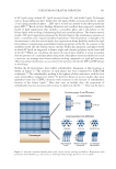
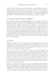
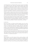
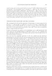
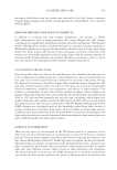
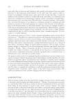
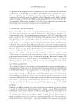
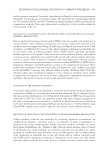
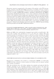
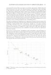
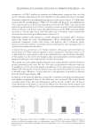
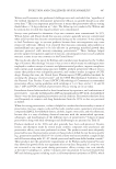
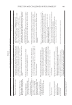
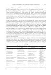
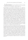
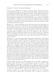
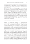
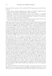
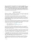
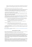
Figure 1. Diagram showing layers of the epidermis. Illustration by Robin M. Wickett.
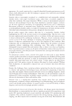
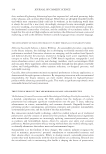
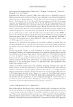
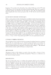
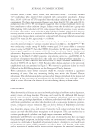
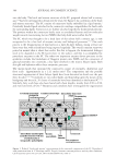
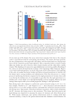
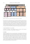
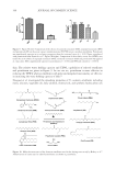
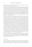
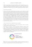
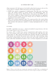
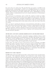
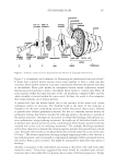
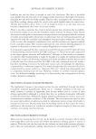
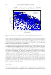
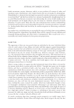
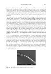
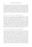
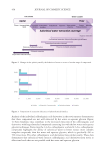
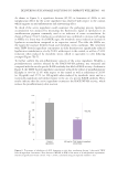
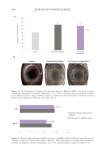
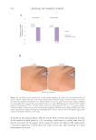
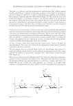
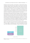
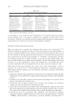
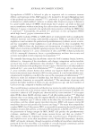
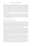
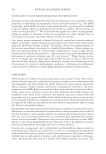
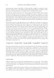
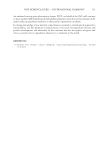
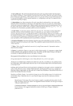
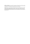
Figure 2. Schematic of the “bricks and mortar” model for human stratum corneum illustrating the corneocyte
“bricks,” the intercellular lipid “mortar” and the desmosomes connecting the corneocytes.
model that includes the presence of the desmosomes that hold SC cells together prior to
desquamation and discusses the role of natural moisturizing factor in SC function.9 This
model is illustrated in Figure 2.
The SC primarily forms in the stratum granulosum (SG), named for the granules that
appear in the cells comprised of keratohyalin granules10 composed of protein and lamellar
bodies that contain lipids.11,12 In the SG: the nucleus is digested, the cytoplasm disappears,
and the lipids that will eventually form the SC barrier are released into the intercellular
space.7,13–15 Epidermal keratins aggregate to form microfibrils16 inside the corneocytes
under the influence of filaggrin from the keratohyalin granules.17–19 The keratinocyte cell
membrane is replaced by the corneocyte cell envelope made of cross-linked protein from
the cytoplasm and keratohyalin granules and lipids that are covalently attached to its
Figure 1. Diagram showing layers of the epidermis. Illustration by Robin M. Wickett.
Figure 2. Schematic of the “bricks and mortar” model for human stratum corneum illustrating the corneocyte
“bricks,” the intercellular lipid “mortar” and the desmosomes connecting the corneocytes.