346 JOURNAL OF COSMETIC SCIENCE
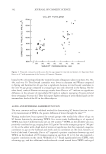
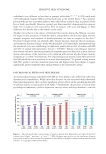
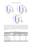
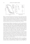
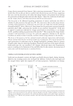
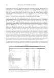
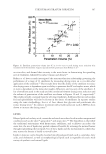
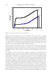
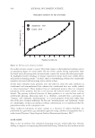
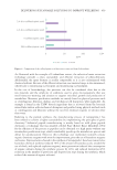
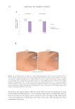
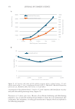
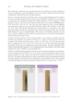
The total time for SC on the volar forearm to turn over increased from 19.8 ± 1.4 days in
the under 35 years old group to 28.1 ± 2.7 days in an over 60 age group.56 Similar changes
were seen on the upper inner arm as shown in Table II. Their results agree with the positive
correlation between age and the time to turn over the SC reported by Roberts and Marks.87
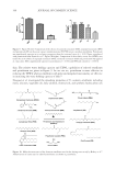
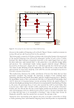
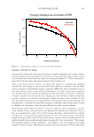
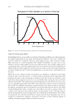
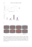
Grove found the number of SC cell layers was approximately constant between age groups
(Table II) indicating that the increased SC turnover time results from decreased epidermal
cell proliferation rates with age.24,56 Marks reported a small but statistically significant
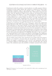
decline with age in the number of epidermal cells in the DNA synthesis phase.88 Engelke
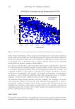
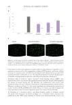
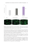
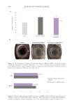
et al. investigated differences between young normal skin, young dry skin, aged normal
skin, and aged dry skin89 using an antibody that stains the nuclei of proliferating cells (Ki-
S3).90 Their data also indicate a significant reduction in epidermal proliferation rate in aged
“normal skin” compared to young normal skin. A slower rate of epidermal cell proliferation
is consistent with the significant increase in time to repair a disrupted SC barrier reported
by Ghadially et al.85 Grove also reported that small chemically induced blisters healed
more slowly in subjects between 65–70 years of age than in subjects 18–25 years of age.91
DRY SKIN IN THE ELDERLY
The greater tendency of older subjects to develop dry skin is well known.4,92–94 Elderly dry
skin is especially prone to itching which can sometimes be severe.48–50
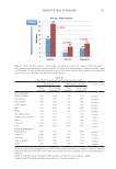
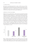
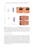
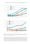
It is not entirely clear why older subjects are more prone to developing dry skin. One
possibility is lower levels of NMF with age as NMF levels are reported to be lower in dry
skin.40 Data on the effect of age on NMF levels do not present a consistent picture. One
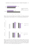
of the key moisturizing components of NMF is PCA.34,40 Harding et al. reported lower
levels of PCA in older subjects and the difference was more marked deeper in the stratum
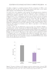
corneum.99 Horii et al. reported a general decrease in free amino acids with increasing
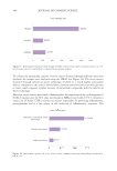
levels of dry skin in elderly subjects.100 On the other hand Jacobson et al. found that some
SC amino acids decreased with age while others increased, but no difference was seen in
total NMF free amino acids (FAA) normalized to protein content between young and old
subjects.101 Takahashi and Tezuka102 reported that NMF FAA increased in older subjects.
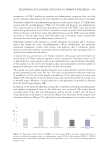
However, both groups sampled the stratum corneum by scraping skin flakes from the
surface rather than tape stripping to sample from lower levels of the SC as Harding et al.
did. Further, as Tagami103 has pointed out Takahashi and Tezuka reported their results
as FAA/corneocyte. Thus, the higher levels of NMF FAA they report could be due to the
presence of larger corneocytes in older skin25,26,56 as discussed above.
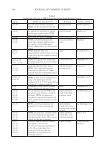
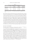
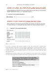
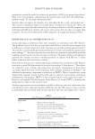
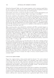
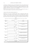
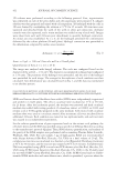
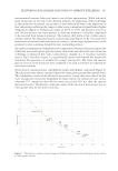
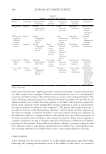
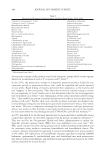
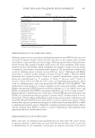
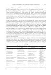
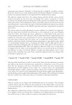
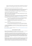
Table II
Measurements of Epidermal Turnover Rate From Grove et. al56
Site Age Transit time
(days)
Number of
cell layers
Turnover rate
(hours/layer)
Volar forearm 35 19.8 ± 1.39 17.0 ± 0.83 28.3 ± 1.2
Volar forearm 60 28.1 ± 2.66 16.8 ± 0.66 40.1** ± 3.8
Upper inner arm 35 17.7 ± 2.02 14.3 ± 0.61 30.0 ± 2.6
Upper inner arm 60 25.5 ± 2.63 13.9 ± 0.81 46.6 ± 6.5
Results are presented as mean ± S.E. Calculated from the data due to a typographical error in
paper.
The total time for SC on the volar forearm to turn over increased from 19.8 ± 1.4 days in
the under 35 years old group to 28.1 ± 2.7 days in an over 60 age group.56 Similar changes
were seen on the upper inner arm as shown in Table II. Their results agree with the positive
correlation between age and the time to turn over the SC reported by Roberts and Marks.87
Grove found the number of SC cell layers was approximately constant between age groups
(Table II) indicating that the increased SC turnover time results from decreased epidermal
cell proliferation rates with age.24,56 Marks reported a small but statistically significant
decline with age in the number of epidermal cells in the DNA synthesis phase.88 Engelke
et al. investigated differences between young normal skin, young dry skin, aged normal
skin, and aged dry skin89 using an antibody that stains the nuclei of proliferating cells (Ki-
S3).90 Their data also indicate a significant reduction in epidermal proliferation rate in aged
“normal skin” compared to young normal skin. A slower rate of epidermal cell proliferation
is consistent with the significant increase in time to repair a disrupted SC barrier reported
by Ghadially et al.85 Grove also reported that small chemically induced blisters healed
more slowly in subjects between 65–70 years of age than in subjects 18–25 years of age.91
DRY SKIN IN THE ELDERLY
The greater tendency of older subjects to develop dry skin is well known.4,92–94 Elderly dry
skin is especially prone to itching which can sometimes be severe.48–50
It is not entirely clear why older subjects are more prone to developing dry skin. One
possibility is lower levels of NMF with age as NMF levels are reported to be lower in dry
skin.40 Data on the effect of age on NMF levels do not present a consistent picture. One
of the key moisturizing components of NMF is PCA.34,40 Harding et al. reported lower
levels of PCA in older subjects and the difference was more marked deeper in the stratum
corneum.99 Horii et al. reported a general decrease in free amino acids with increasing
levels of dry skin in elderly subjects.100 On the other hand Jacobson et al. found that some
SC amino acids decreased with age while others increased, but no difference was seen in
total NMF free amino acids (FAA) normalized to protein content between young and old
subjects.101 Takahashi and Tezuka102 reported that NMF FAA increased in older subjects.
However, both groups sampled the stratum corneum by scraping skin flakes from the
surface rather than tape stripping to sample from lower levels of the SC as Harding et al.
did. Further, as Tagami103 has pointed out Takahashi and Tezuka reported their results
as FAA/corneocyte. Thus, the higher levels of NMF FAA they report could be due to the
presence of larger corneocytes in older skin25,26,56 as discussed above.
Table II
Measurements of Epidermal Turnover Rate From Grove et. al56
Site Age Transit time
(days)
Number of
cell layers
Turnover rate
(hours/layer)
Volar forearm 35 19.8 ± 1.39 17.0 ± 0.83 28.3 ± 1.2
Volar forearm 60 28.1 ± 2.66 16.8 ± 0.66 40.1** ± 3.8
Upper inner arm 35 17.7 ± 2.02 14.3 ± 0.61 30.0 ± 2.6
Upper inner arm 60 25.5 ± 2.63 13.9 ± 0.81 46.6 ± 6.5
Results are presented as mean ± S.E. Calculated from the data due to a typographical error in
paper.