348 JOURNAL OF COSMETIC SCIENCE
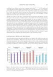
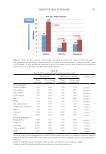
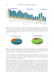
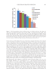
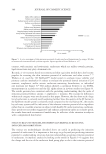
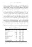
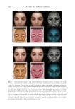
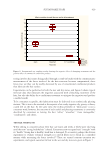
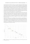
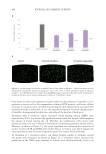
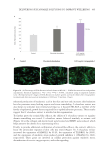
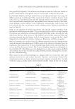
TEWL with age on most body sites (Table 1 and Akdeniz et al.77) but while statistically
significant the observed TEWL reductions may not be clinically relevant as pointed out
above. It also appears that the immediate response of the SC barrier to applied irritants is
not greater in older skin.
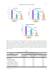
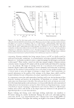
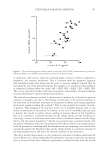
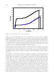
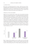
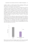
One result that is likely to be of clinical significance is the significantly reduced rate of SC
barrier repair after disruption reported by Ghadially et al.85 This effect is possibly due to
the reduced turnover rate of the SC24,56 as discussed above. So, while the SC barrier may not
be easier to disrupt in elderly skin it is slower to heal once disrupted.
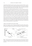
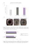
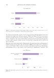
While the tendency of older subjects to develop dry skin is well known, the source of this
problem is not yet completely clear and deserves further study.
REFERENCES
(1) Lavker RM, Zheng PS, Dong G. Morphology of aged skin. Clin Geriatr Med. 1989 5:53–67.
(2) Gilchrest BA. Skin aging and photoaging: an overview. J Am Acad Dermatol. 1989 21:610–613.
(3) Kohl E, Steinbauer J, Landthaler M, Szeimies RM. Skin ageing. J Eur Acad Dermatol Venereol.
2011 25:873–884.
(4) Wickett RR, Tate M. Part 3.2.4 Compromised Skin in the Elderly. In: Rosen M, ed. Harry’s Cosmeticology.
9th ed. Volume 1. Gloucester, MA: Chemical Publishing Company Inc 2015:282–328.
(5) Madison KC. Barrier function of the skin: “la raison d’être” of the epidermis. J Invest Dermatol.
2003 121:231–241.
(6) Elias PM, Choi EH. Interactions among stratum corneum defensive functions. Exp Dermatol.
2005 14:719–726.
(7) Elias PM. Epidermal lipids, barrier function, and desquamation. J Invest Dermatol. 1983 80:44–50.
(8) Talreja P, Kleene N, Pickens W, Wang T, Kasting G. Visualization of the lipid barrier and measurement
of lipid pathlength in human stratum corneum. AAPS PharmSci J. 2001 3
(9) Harding CR. The stratum corneum: structure and function in health and disease. Dermatol Ther. 2004 17
Suppl 1:6–15.
(10) Odland GF. Structure of the skin. In: Goldsmith LA, ed. Physiology, Biochemistry and Molecular Biology of
the Skin. 2nd ed. New York, NY: Oxford University Press 1991:3–62.
(11) Odland GF. A submicroscopic granular component in human epidermis. J Invest Dermatol. 1960 34:11–15.
(12) Oashi M, Sawada Y, Makita R. Odland body and intercellular substances. Acta Derm Venereol Suppl
(Stockh). 1973 73:47–54.
(13) Elias PM, Grayson MA, Lampe MA, Williams ML, Brown BE. The Intercorneocyte Space. In: Marks R,
Plewig G, eds. Stratum Corneum. New York, NY: Springer-Verlag 1983:53–67.
(14) Elias PM, Menon GK. Structural and lipid biochemical correlates of the epidermal permeability barrier.
Adv Lipid Res. 1991 24:1–26.
(15) Wertz PW, Downing DT. Glycolipids in mammalian epidermis: structure and function in the water
barrier. Science. 1982 217:1261–1262.
(16) Steinert PM, North AC, Parry DA. Structural features of keratin intermediate filaments. J Invest
Dermatol. 1994 103:19S–24S.
(17) Dale BA, Holbrook KA, Kimball JR, Hoff M, Sun TT. Expression of epidermal keratins and filaggrin
during human fetal skin development. J Cell Biol. 1985 101:1257–1269.
(18) Dale BA, Presland RB, Lewis SP, Underwood RA, Fleckman P. Transient expression of epidermal
filaggrin in cultured cells causes collapse of intermediate filament networks with alteration of cell shape
and nuclear integrity. J Invest Dermatol. 1997 108:179–187.
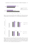
TEWL with age on most body sites (Table 1 and Akdeniz et al.77) but while statistically
significant the observed TEWL reductions may not be clinically relevant as pointed out
above. It also appears that the immediate response of the SC barrier to applied irritants is
not greater in older skin.
One result that is likely to be of clinical significance is the significantly reduced rate of SC
barrier repair after disruption reported by Ghadially et al.85 This effect is possibly due to
the reduced turnover rate of the SC24,56 as discussed above. So, while the SC barrier may not
be easier to disrupt in elderly skin it is slower to heal once disrupted.
While the tendency of older subjects to develop dry skin is well known, the source of this
problem is not yet completely clear and deserves further study.
REFERENCES
(1) Lavker RM, Zheng PS, Dong G. Morphology of aged skin. Clin Geriatr Med. 1989 5:53–67.
(2) Gilchrest BA. Skin aging and photoaging: an overview. J Am Acad Dermatol. 1989 21:610–613.
(3) Kohl E, Steinbauer J, Landthaler M, Szeimies RM. Skin ageing. J Eur Acad Dermatol Venereol.
2011 25:873–884.
(4) Wickett RR, Tate M. Part 3.2.4 Compromised Skin in the Elderly. In: Rosen M, ed. Harry’s Cosmeticology.
9th ed. Volume 1. Gloucester, MA: Chemical Publishing Company Inc 2015:282–328.
(5) Madison KC. Barrier function of the skin: “la raison d’être” of the epidermis. J Invest Dermatol.
2003 121:231–241.
(6) Elias PM, Choi EH. Interactions among stratum corneum defensive functions. Exp Dermatol.
2005 14:719–726.
(7) Elias PM. Epidermal lipids, barrier function, and desquamation. J Invest Dermatol. 1983 80:44–50.
(8) Talreja P, Kleene N, Pickens W, Wang T, Kasting G. Visualization of the lipid barrier and measurement
of lipid pathlength in human stratum corneum. AAPS PharmSci J. 2001 3
(9) Harding CR. The stratum corneum: structure and function in health and disease. Dermatol Ther. 2004 17
Suppl 1:6–15.
(10) Odland GF. Structure of the skin. In: Goldsmith LA, ed. Physiology, Biochemistry and Molecular Biology of
the Skin. 2nd ed. New York, NY: Oxford University Press 1991:3–62.
(11) Odland GF. A submicroscopic granular component in human epidermis. J Invest Dermatol. 1960 34:11–15.
(12) Oashi M, Sawada Y, Makita R. Odland body and intercellular substances. Acta Derm Venereol Suppl
(Stockh). 1973 73:47–54.
(13) Elias PM, Grayson MA, Lampe MA, Williams ML, Brown BE. The Intercorneocyte Space. In: Marks R,
Plewig G, eds. Stratum Corneum. New York, NY: Springer-Verlag 1983:53–67.
(14) Elias PM, Menon GK. Structural and lipid biochemical correlates of the epidermal permeability barrier.
Adv Lipid Res. 1991 24:1–26.
(15) Wertz PW, Downing DT. Glycolipids in mammalian epidermis: structure and function in the water
barrier. Science. 1982 217:1261–1262.
(16) Steinert PM, North AC, Parry DA. Structural features of keratin intermediate filaments. J Invest
Dermatol. 1994 103:19S–24S.
(17) Dale BA, Holbrook KA, Kimball JR, Hoff M, Sun TT. Expression of epidermal keratins and filaggrin
during human fetal skin development. J Cell Biol. 1985 101:1257–1269.
(18) Dale BA, Presland RB, Lewis SP, Underwood RA, Fleckman P. Transient expression of epidermal
filaggrin in cultured cells causes collapse of intermediate filament networks with alteration of cell shape
and nuclear integrity. J Invest Dermatol. 1997 108:179–187.