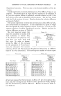
182 JOURNAL OF THE SOCIETY OF COSMETIC CHEMISTS noted, but effectively used in dermatologic therapy. Patients suffering from dermatoses characterized by excessive amounts of dry scaling and excessive dryness of the horny surfaces (e.g., ichthyosis, palmar and plantar hyperkeratosis, ichthyosiform and certain exfoliative erythrodermas) have, since decades and whenever feasible, been sent to climates with obtaining high temperatures and high relative .humidities--and very often do quite well there. Contrariwise, patients with excessively oily and greasy skin surfaces or with skin diseases connected with an excess of the aqueous and oily surface film (-e.•.•ebor•hea. rae•e. vulgaris), often do poorly in hot humid climates, but well in hot dry environments. These phenomena will be discussed somewhat further when I take up more details dealing with the clinical implications of the laboratory data. But at this point I must not fail to add that the softening of the keratinous skin surface in a moist environment need by no means be entirely due to the lipid moiety of the surface emulsion but can, and perhaps in great measure, be attributed to the watery component. For as has been em- phasized by the beautiful experiments of Blank and associates, hydrated keratin is soft and pliable while dehydrated keratin is hard and brittle (37). Of all the laboratory findings which speak in favor of the great role of thermogenic sweat in aiding the delivery and accelerating the spread of skin surface lipids, the following are to our minds among the most con- vincing: First our numerous experimental observations showing that short exposure of a subject to gradually rising temperatures produces an in- crease in the amount of surface lipids as soon as the particular skin area under study begins to show visible sweating. With the one exception of the axillary vaults, in all the areas we studied we noted the synchronous appearance of visible sweat and a sharp rise in quantity of surface lipids (21,23). The results of another of our clinical investigations speak in exactly the same direction. In three of our patients with emotional hyperhidrosis* * In these patients the parallel increase of sweat and oflipids was demonstrated on the palms --on which emotional hyperhidrosis is usually most strongly manifest, and which, with the soles, are often the only areas affected. Since the palms have no sebaceous glands, the source of the demonstrated lipids here requires explanation. At this point one must therefore recall that the lipid phase of the skin surface emulsion is not derived from the sebaceous glands alone but also from epidermal cells as a result of their disintegration and transformation into kera- tin. The fraction derived from the epidermal cells, the so called "horn-fat," or better "horn- wax," constitutes a relatively small proportion of the lipid film in all those skin areas in which sebaceous glands are present and deliver by far the larger portion. The horn-wax contains considerably more steroids, in particular more cholesterol, and less squalene than sebum and in contrast to sebum hardly any triglycerides. As we have shown by systematic carefully controlled studies of the palmar surfaces (where, as stated, sebaceous glands are absent, but sweat glands present in abundance) the appearance of sweat droplets is regularly associated with the appearance of horn-wax as well (23). We believe that the horn-wax may well be largely responsible for the close quantitative correlation between the aqueous and lipid phase, which, as described, exists practically all over the body surface.

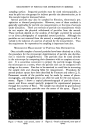
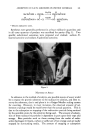
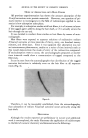
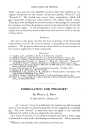
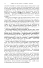
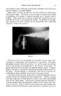
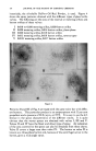
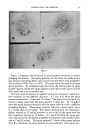
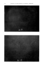
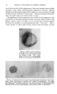
CLINICAL DISTURBANCES IN SWEATING 183 in which the sweat poured out without any increase in environmental or skin surface temperatures, we could demonstrate that the skin lipids and their rate of delivery increased in parallel with this nonthermogenic increase in sweating. Studies by Direct Observations ( Fisualization, etc.) of the Delivery, Emulsifica- tion and Spread of the Sweat and Lipids of the Skin's Surface Many observers using a variety of techniques have directly observed the appearance and spread of the surface film upon skin areas which have first been cleansed. Thus Butcher and his group observed a rather quick reappearance of the surface lipids and their spread through the skin sulci (32). As stated, contrary to our own concept, Butcher and co-workers attrib- uted these phenomena to a lowering of the viscosity of the lipids themselves rather than to the role of sweat as an emulsifier. However, Jones and collaborators demonstrated that the lipids spread over the skin surface at the rate of 3.3 cm. per second when that surface is moistened by sweat but that they do not spread at all, or only very slowly when the skin has first been cleansed with ether or alcohol (24). Similarly, or rather in a complementary fashion, Schneider and Schuleit demonstrated a con- siderable reduction in the spread of water on the skin surface and an in- crease of interface tension between water and surface after previous cleans- ing of the skin surface with alcohol, ether, alcohol plus ether or even water alone. And completing the chain of evidence, the last-named investigators showed that when the lipid materials which had been removed with these solvents (and in particular those removed with alcohol) were reapplied to the cleansed skin areas, or even applied to previously uncleansed skin areas, the wetting of the skin surface by water was greatly facilitated (25). While all these findings speak strongly for the reciprocal emulsification of the ether-soluble materials and of sweat on the skin surface, our own studies enabled us to see these phenomena actually taking p/ace. In these studies we employed a binocular wide angle surface reflectance microscope and a light source which enabled us clearly to see the skin's surface at relatively great magnification (23). In the following I shall call this technique "skin surface microscopy." In numerous experiments we exposed skin areas (which had preliminar'ily been cleansed with ether) to the vapors of osmic acid under strictly comparable and controlled con- ditions. The ensuing black coloration produced by the reduction of osmic tetroxide in the presence of lipid components was observed and localized by skin surface microscopy. In addition to the characteristic darkening of the follicular ostia, we saw a consistent and unmistakable blending of sweat and lipids on the skin's surface, especially after provocation of thermal sweating. About
Purchased for the exclusive use of nofirst nolast (unknown)
From: SCC Media Library & Resource Center (library.scconline.org)