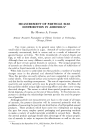
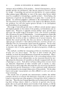
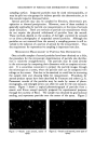
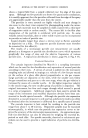
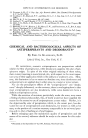
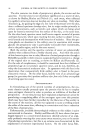
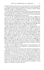
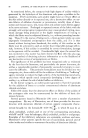
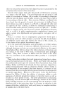
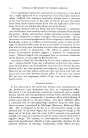
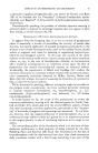
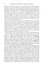
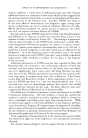
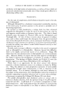
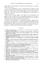
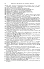
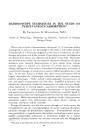
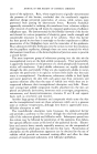
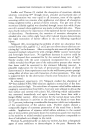
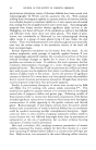
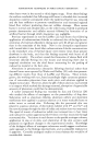
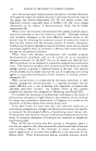
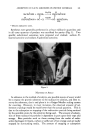
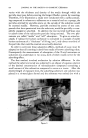
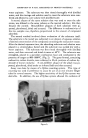
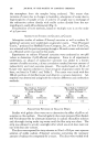
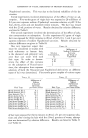
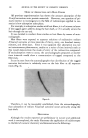
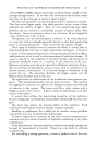
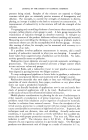
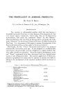
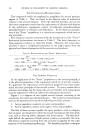
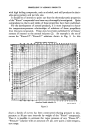
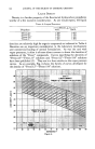
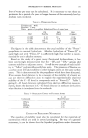
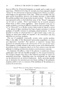
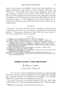
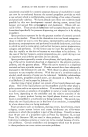
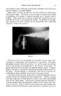
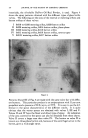
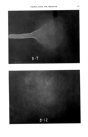
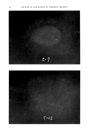
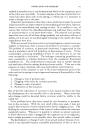
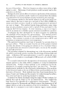
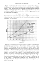
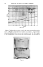
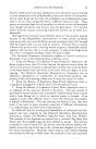
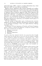
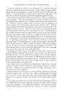
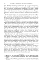
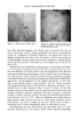
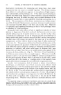
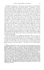
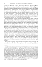
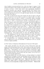
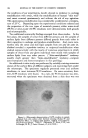
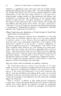
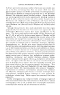
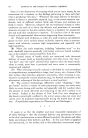
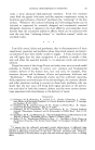
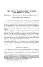
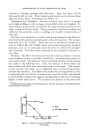
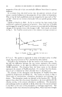
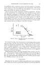
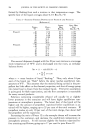
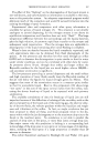
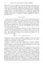
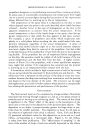
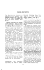
184 JOURNAL OF THE SOCIETY OF COSMETIC CHEMISTS seven minutes after the first appearance of the sweat droplets many of these assumed a dark shade which gradually deepened in intensity. Shortly thereafter the black, glistening emulsion began to travel and spread through the fine network of the skin's surface sulci. Maximal staining of the entire skin area under study was reached within an hour. The significance of the outpouring sweat droplets for the appearance and distribution of the blackened lipid material was most clearly visible on the palmar surfaces (where the lipids are not derived from sebaceous glands but from the cornified or cornifying epidermal cells--"horn-wax." After a "latent period" of about eight minutes during which the freshly secreted Figure 4.--Palmar surface exposed to osmic acid vapors. Dark sweat droplets in "Beakers of Sweat Secretion" and dark material in sulci. (9X) (Herr- mann, F., Prose, P. H., and Sulzberger, M. B., •. Invest. Dermat., 21, 403 (1953).) Figure 5.--Symmetrical skin areas exposed to osmic acid vapors after thermal sweat stimu- lation. Right: Atropinized site (atropine sulfate applied by iontophoresis). Very slight darkening, essentially confined to follicular ostia. Left: Control site (distilled water applied by iontophoresis). Site intensely darkened. Sweat droplets visible on and around this area (•Hermann, F., Prose, P. H., and Sulzberger, M. B., •e. Invest. Dermat. 21 406 (1953).)

CLINICAL DISTURBANCES IN SWEATING 185 sweat droplets remained perfectly clear, dark dots began to appear in the centers of the bases of the cup-like sweat gland ostia, the so-called "beakers of sweat secretion." From these points dark pseudopodia (Schlieren) gradually extended both upwards and peripherally until finally every sweat droplet was uniformly black. Rapidly thereafter, the tar-like mass moved through the skin sulci, first through the larger channels, then through the smaller ones (Fig. 4). In order to demonstrate even more clearly the role of sweat in the de- livery and spread of surface lipids we injected attopine locally into ether- cleansed skin areas (in order to inhibit or materially reduce sweating). After general thermal stimulation of sweating, we exposed these atropin- ized areas, as well as symmetrically situated nonatropinized areas, to osmic acid vapors, and by skin surface microscopy, observed the differences in the appearance and distribution of lipids. In these experiments, the surface of the atropinized areas invariably remained much lighter than that of the nonatropinized control areas (Fig. 5). Moreover, following the appearance of sweating, a third skin site was carefully wiped with dry muslin (to remove the surface sweat and lipid emulsion) and then immedi- ately exposed to osmic acid vapors. Under these conditions the nonatro- pinized wiped control site showed little if any more darkening than the atropinized site. From these results we drew the following inferences: 1. The darkening of the skin surface produced by osmic acid vapors is due principally to the uppermost film, the "surface lipids" which are present on the upper surface of, and external to, the stratum comeurn. 2. Atropinization, i.e., decreased outpouring of sweat, interferes with the appearance of the outer lipid film--in agreement with our concept of sweat as lipid emulsifier. In Ifitro Studies on Reciprocal Emulsification of Sweat and Skin Lipids The studies described up to this point concern observations upon the surface of the living human skin in situ their results are thus quite directly applicable to the actual clinical and cosmetic problems of dealing with the human skin. Nevertheless, we felt that additional information might be obtained by in vitro studies of actual sweat and of actual ether-soluble material which had been removed from the skin surface, and carried out a series of such in vitro experiments. By using suitable dyes we could observe a strong tendency to mutual emulsification of samples of sweat and the lipid material collected by ether washings from the skin's surface (23). All sweat samples tested were much more efficient in this regard than water. Moreover, the tendency of the human skin surface lipids to undergo emulsification with sweat, or even with water, was incomparably stronger than that of lanolin. Indeed under
Purchased for the exclusive use of nofirst nolast (unknown)
From: SCC Media Library & Resource Center (library.scconline.org)