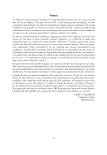
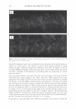
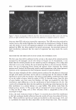
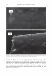
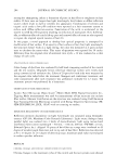
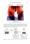
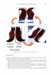
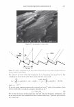
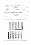
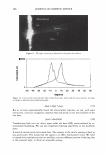
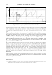
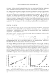
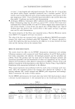
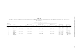
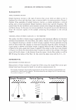
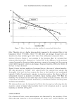
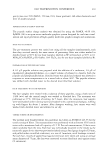
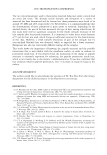
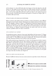
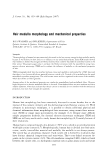
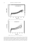
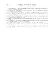
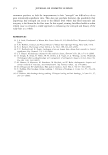
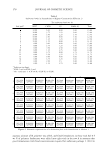
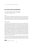
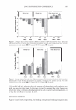
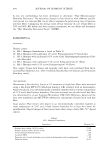
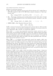
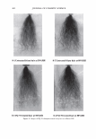
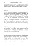
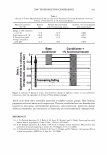
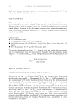
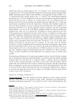
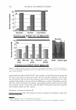
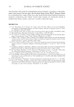
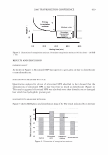
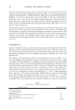
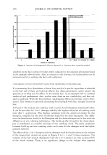
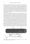
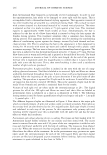
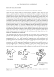
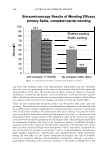
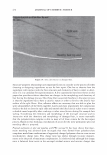
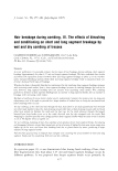
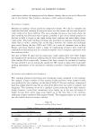
310 ': 1 JOURNAL OF COSMETIC SCIENCE aml -2.8 -3 -3.2 f----+-----+,o--- ,s,------ 20 -,.--1- -----'- --+25-- -----35 J0 +-----+-......,_40 __ aml Path length - I Figure 1. Surface roughness profile of a European Brown hair fiber. requires a metal coating on the sample this enables the high vacuum mode of the SEM to be used during image capture. A previous study proved that coating with a thin layer of metal had no measurable effect on the depth information produced by this method [l}. The second protocol analyses the same area of a single fiber before and after treatment. An area to be analyzed is firstly selected and the surface profile is created using the SEM and MeX technique. This fiber can then be treated and then the same area located using a co-ordinate system and another surface profile can be created. This enables us to directly compare the surface profile of the hair fiber before and after a treatment cycle. As a direct comparison is made it is not possible to coat the hair fibres so the variable pressure mode of the SEM is used. In order to validate the novel technique and ensure that the scale heights measured were indeed accurate and reproducible, extensive calibration of the SEM and the 3D software has been performed. In addition scale heights on a single hair have been determined using Atomic Force Microscopy (AFM) and the results compared with analogous data produced from the same scale edges using the 3-D image analysis technique. When comparing these scale edges using the two methods it demonstrated that there is minimal variation in the measured scale heights using this novel 3-D image analysis technique The work done indicates that this is a quick, accurate and viable method to determine scale height in keratin fibers. This method has enabled us to analyze the effect of various treatments applied to hair fibers. REFERENCES (1) C. Tomes, J. T. Jones, C. M. Carr, and D. Jones, Three-dimensional imaging anl analysis of the surface of hair fibers using scanning electron microscopy, J. Cosrnet. Sci. (in press).

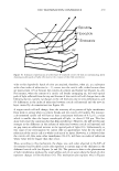
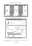
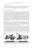
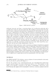
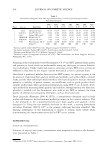
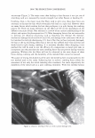
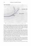
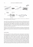
]. Cosmet. Sci. 1 58, 311-317 Quly/August 2007) Fundamental DSC investigations of a-keratinous materials as basis for the interpretation of specific effects of chemical, cosmetic treatments on human hair F.-J. WORTMANN, G. SENDELBACH, and C. POPESCU, University of Manchester, School of Materials, P.O. Box 88, Manchester M60 JQD, UK (F.-j. W.), Wella Service GmbH, Berliner Allee 65, D-64274 Darmstadt, Germany (G.S.), and DWI at RWTH Aachen University, Pauwelsstrasse 8, D-52074 Aachen, Germany (C.P). Synopsis a-keratinous materials can be considered as two-phase, filament/matrix composites, in which partly crys- talline, a-helical intermediate filaments (IF) are embedded in an amorphous matrix of IF-associated proteins (IFAP). Differential Scanning Calorimetry (DSC) of keratins in water was found to be especially suited to analyze various aspects of the thermal stability of these main morphological components. Results and considerations are reviewed, which were gained by applying the principles derived from fundamental investigations to the specific effects of oxidation (bleaching) and reduction (perm-waving). Properties and interactions of the main morphological components of human hair are considered that are specifically related to the various aspects of their thermal stability. The overall view of the results shows that the course of the thermal unfolding of the a-helix in the IFs is independent of the chemical history of hair. The matrix properties are the primary factor controlling the kinetics of the onset of the denaturation process in the IF/IF AP-composite. INTRODUCTION Hair, such as animal or human hair, grows in various types on all parts of the body and from cavities in the skin, called follicles, which are embedded up to 3 mm deep in the dermis and extend to the surface of the skin through the epidermis and the stratum corneum (1). During its growth the hair fiber, in the same way as other a-keratin materials, such as hoof, horn, and quill, develops complex morphological fine structures (2). Figure 1 shows a graphical representation of the structure of a wool fiber, as possibly the best investigated a-keratin hair fiber (3 ,4). A hair fiber is constituted of cells that differentiate during hair growth to form, namely, the fiber core (cortex) and an outer protective layer (cuticle). The interface between the Presented in specific part at the 34th Annual Conference of the North American Thermal Analysis Society, August 2006, Bowling Green, KY. Address all correspondence to F.-J. Wortmann. 311
Purchased for the exclusive use of nofirst nolast (unknown)
From: SCC Media Library & Resource Center (library.scconline.org)