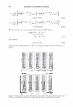
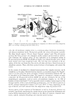
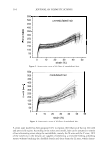
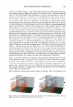
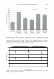
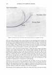
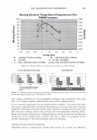
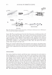
360 JOURNAL OF COSMETIC SCIENCE (2% w/w cystine) (10,11). CMC and endocuticle are believed to be the main diffusion pathways into human hair (12,13). The cortex is formed by elongated cortical cells surrounded by the CMC. The cortical cell (-100 µm long and 3 µm wide) has an inner fibrillar structure (macrofibrils of 0.4 µm diameter formed by the intermediate filaments of -10 nm diameter) embedded in a hydrophilic sulphur-rich matrix (12). This well-defined structure gives rise to out- standing mechanical properties (14). The medulla is located in the centre of the fibre and may be absent, fragmented or continuous (15). It is amorphous and has a high lipid content com pared to the rest of the fibre (16). Morphologically, it has been reported that medulla has a porous structure formed by "spongy" keratin (17 ,18) and some vacuoles filled with air resulting from the differentiation process (17, 19). A layer of CMC separates the medulla from the cortex (20). Nevertheless, some authors say that the medulla is just those vacuoles or granules and that the porous structure is formed by an unidentified material (17 ,20). Medulla represents about 20% of the total fibre section. The effects of this porous structure on mechanical properties are still unknown. The cuticle subunits were first studied by TEM (21). In TEM, the contrast is created by staining the sliced sample to obtain a two dimensional image. Human hair has to be embedded in a resin to be sliced in an ultramicrotome, otherwise the ultrastructure is not differentiated. In the case of medulla observations, preserving the porous structure and the CMC layer is also difficult. This paper aims to identify the microfibrils in the medulla by TEM and relate their structures to the mechanical properties. MATERIALS AND METHODS A one-head tress was observed in a stereoscope and separated into bundles of 40 fibers with or without medulla. HAIR SAMPLES A Caucasian dark brown hair sample was obtained from a female donor and separated in medullated and unmedullated fibers using a stereo-microscope. This procedure elimi- nates chemical and morphological differences by genetics or cosmetic treatments since all fibers come from the same scalp. TEM OBSERVATIONS TEM micrographs were obtained in a Philips CM200 microscope, operating at 160 kV. One cm segments of hair fibers were fixed using 2 ml of 2% (v/v) OsO4 (Sigma) in 0.1 mol L - 1 sodium cacodilate buffer at pH 7, for 4 h in the dark, followed by washing in water or buffer for 30 min. The segments were dehydrated with a series of ethanol solutions of increasing concentration (from 50 to 100%, v/v), for 15 min, two times with each one of the solutions. Then, the hair was exposed twice to a solution of 100% ethanol for 5 min. Spurr resin (standard formulation of low hardening rate, 0.2 g catalyst) used as embedding media, in the following steps: (a) hair segments were placed in closed

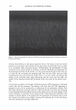
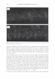
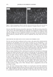
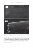
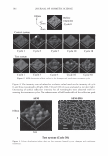
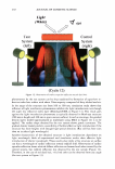
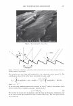
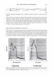
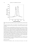
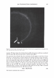
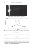
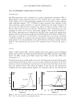
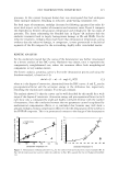
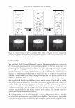
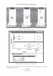
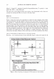
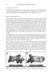
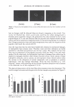
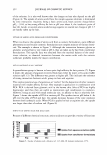
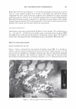
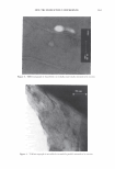
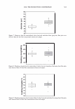
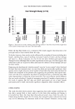
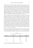
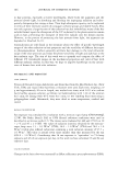
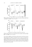
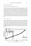
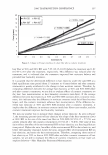
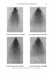
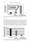
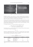
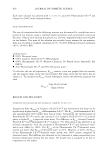
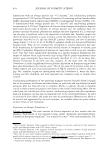
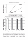
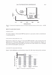
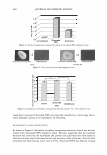
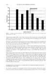
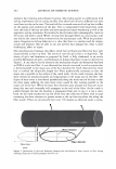
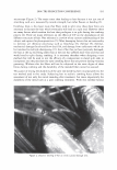
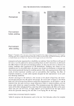
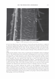
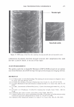
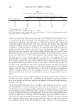
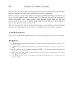
2006 TRI/PRINCETON CONFERENCE 361 flasks filled with resin and ethanol (1: 1, v/v) solution, (b) flasks were placed in an acrylic rotor at 3 rpm constant rotation for 4-8 days, (c) the flasks were opened for ethanol evaporation for 24 h, and (d) the hair segments were transferred to proper inclusion molds and cured at 70°C for 24 h. Ultra-thin sections were cut using a Sorvall Porter- Blum Mt2-B ultramicrorome, mounted in a 200 mesh grid and stained with a freshly prepared aqueous solution of 2% uranyl acetate for 1 S min and 1 % lead citrate for 8 min. Samples were stained just few hours before the observations. MECHANICAL PROPERTIES Stress/strain curves were obtained from 40 fibers (5 .0 cm length 24 h conditioning at 25 ± 2°C and SO ± 5% RH) of each sample using a universal testing machine (EMIC DL 2000) with a 10 N load cell operating at 10 mm/min constant speed. The diameter of each fiber was measured after conditioning using a micrometer (Mitutoyo Ltd.). RESULTS AND DISCUSSION MEDULLA MORPHOLOGY BY TEM Figure 1 shows a representative micrograph of medulla using TEM. It is possible to observe a porous structure composed of cortical cells and air filled vacuoles. (21) The cortical cells are deformed or elongated, showing that they are distributed randomly throughout the medulla. The vacuoles are connected to these cells by peptide bonds derived from citrulline residues, according to Clement et al. (22). There are some cavities Fi g ure 1. TEM micrograph of human hair medulla.
Purchased for the exclusive use of nofirst nolast (unknown)
From: SCC Media Library & Resource Center (library.scconline.org)