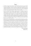
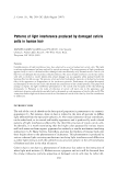
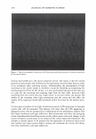
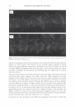
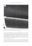
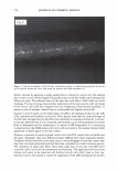
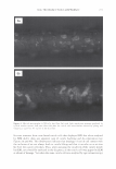
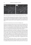
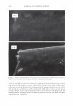
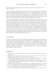
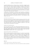
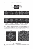
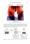
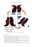
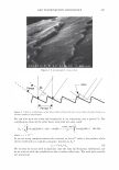
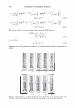
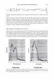
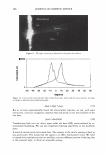
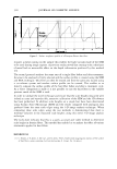
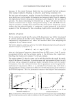
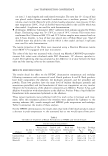
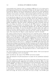
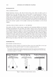
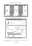
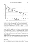
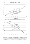
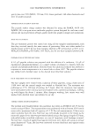
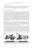
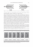
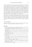
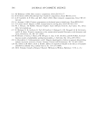
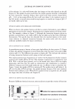
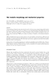
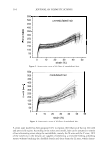
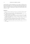
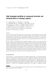
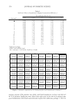
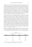
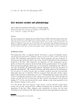
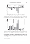
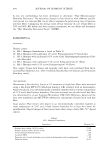
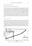
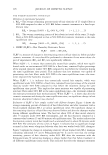
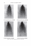
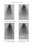
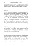
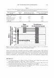
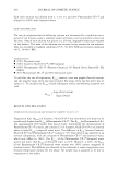
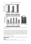
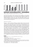
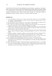
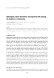
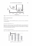
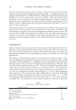
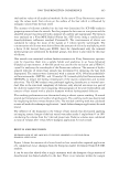
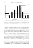
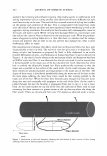
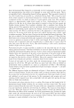
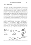
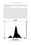
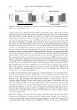
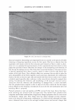
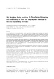
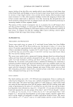
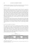
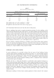
272 JOURNAL OF COSMETIC SCIENCE Figure 3. Optical micrographs (x370) of hair fibers showing strong patterns of light interference after the application of cyclical tension stresses. observed in damaged cuticle cells is considered to be a distortion of the natural iridescent patterns in virgin cuticles cells. Figures 2a and 26 show typical examples of strong patterns of light interference produced by damaged cuticle cells. In Figures 2c and 2d are shown SEM micrographs of the same areas and it can be observed that the cuticle cells have undergone de-cementation and buckling after the application of cyclical extension stresses. The most salient features in the LIPs were their colors and shapes. Commonly observed colors were blue, green, magenta, red, yellow, and white. Most frequently the colors appeared in lines, either, very thin or wide, whose shape was either hyperbolic or straight (see Figures 3a and 36). Other colored patterns that appeared in the form of localized dots (see Figures 4a and 46), clusters of colored dots, and as narrow and long channels were also observed (see Figure 5). Most of these punctual patterns were produced after the hair was subjected, either, to cyclical thermal stresses of wetting and blow drying or to torsion (12-13). It was observed that many of the dot-like patterns tended to dis- appear after the hair was soaked in water for 5 minutes, however, after the fiber was soaked in IPA for 3 minutes the colored dot patterns reappeared again. In many cases similar punctual patterns were observed in hair fibers obtained from common individu- als after the fibers were immersed in IPA for 1 or 2 minutes. In general, the color and shape of the LIPs were dependent on the form in which the

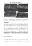
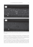
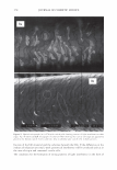
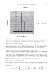
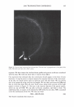
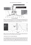
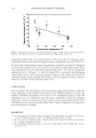
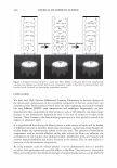
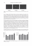
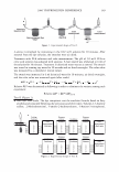
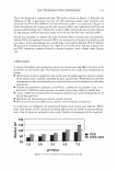
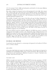
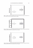
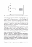
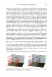
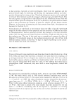
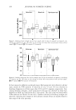
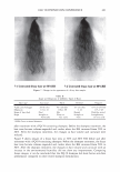
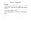
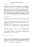
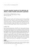
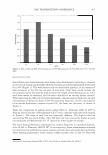
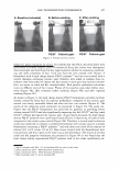
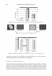
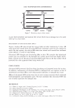
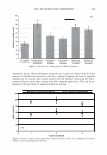
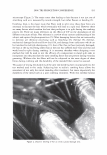
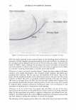
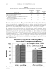
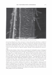
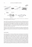
2006 TRI/PRINCETON CONFERENCE 273 Figure 4. Optical (4a) and SEM (46) micrographs (x370) showing letails of dot like patterns appearing in cuticle cells after drying the hair fiber with a hot iron surface at T = 90 C for 5 minutes. Lines and circles in captions help to identify same cuticle sheath areas in both micrographs. cuticle cells were damaged. For instance, the LIPs of hyperbolic shape were seen invari- ably associated to the folding of cuticle cells in buckles. Figures 2a, 26, 2c, and 2d show the juxtaposition of microscopic images displaying the same area of a hair surface with de-cemented and buckled cuticle cells, one obtained by optical microscopy and the other by SEM, respectively. In these figures it can be seen that each cuticle cell that appears folded forming buckles in the SEM micrograph (see Figures 2a and 26) has a corre- sponding hyperbolic pattern of light interference in the picture obtained by optical microscopy (see Figures 2c and 2d). The hyperbolic LIPs were found to be very sensltlve to the application of additional mechanical or swelling stresses to the cuticle cells. Their color and shape underwent dramatic changes when their corresponding cuticle cells were further stressed. Figures 6a and 66 show before and after pictures of a de-cemented and buckled cuticle cell that was
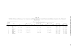
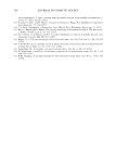
Purchased for the exclusive use of nofirst nolast (unknown)
From: SCC Media Library & Resource Center (library.scconline.org)