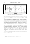
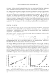
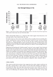
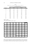
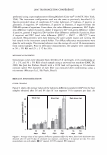
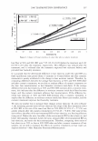
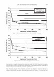
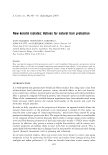
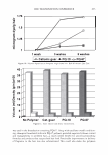
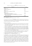
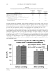
2006 TRI/PRINCETON CONFERENCE 341 purity was over 70% (HPLC, 220 nm, Cl8, linear gradient). All other chemicals used were of analytical grade. PEPTIDE SURFACE CHARGE ANALYSIS The peptide surface charge analysis was obtained by using the PyMOL v0.99 (16). PyMOL (16) is an open source molecular graphics system designed for real time visual- ization and rapid generation of high-quality molecular graphics images and animations. HAIR PRE-TREATMENT The pre-treatment process was carried out using all the samples simultaneously, such that they received exactly the same extent of processing. Hair was either washed in distilled water at 50°C for one hour (samples labelled as W) or bleached, at 50°C in 0.1 M Na 2 CO/NaHCO 3 pH 9 buffer, 10% H 2 O 2 ' also for one hour (samples labelled as B). PREPARATION OF PEPTIDE SOLUTIONS A 0.2 g/1 peptide solution was prepared with the addition of a surfactant, 1.8 g/1 of dipalmitoyl phosphatidylcholine, in a small volume of ethanol to dissolve both the peptides and phosphatidylcholine. Distilled water was added and ethanol was allowed to evaporate at room temperature or in a water bath at about 40°C. The peptide solution was refilled with distilled water to the desired level (the final volume). HAIR TREATMENT WITH THE PEPTIDES The hair samples were treated with a solution of these peptides, using a bath ratio of 1/100 (w/v) and the control sample was washed or bleached hair. The treatment was performed at 3 7°C, 100 rpm of stirring, for 5 hours. After the treatment, hair samples were well washed under running water and washed with a commercial shampoo, rubbing up with fingers for about 1 minute. After shampoo washing, hair tresses were well washed with distilled water and allowed to air dry. TENSILE STRENGTH MEASUREMENTS The method used broadly follows the guidelines laid down in ASTM Dl445-95 for the tensile testing of fibres. The measurements were performed with an Instron 4505 tensile tester with a maximum load cell capacity of 2.5 N. For each measurement, 20 hairs were taken randomly from the tress. Each hair was individually mounted in the tensile jig by means of a paper device that was previously slashed using a fixed gauge length of 20 mm, and pulled under controlled conditions, at a rate of lmm/min, until breakage occurred. For each hair, records of applied load against extension were taken and using an average mean diameter of 7 5 µm, the data were converted to stress (load/unit area) against strain (% extension).

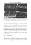
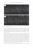
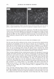
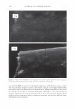
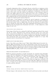
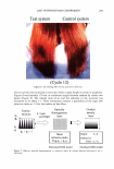
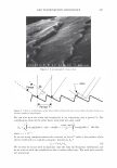
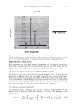
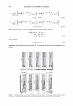
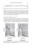
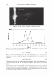
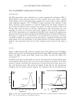
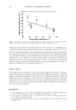
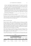
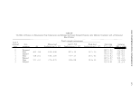
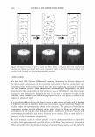
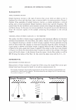
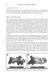
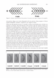
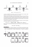
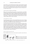
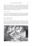
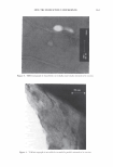
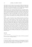
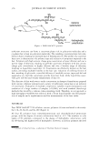
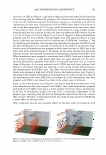
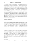
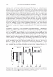
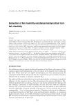
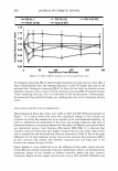
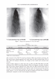
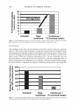
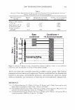
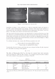
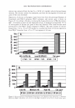
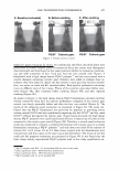
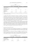
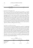
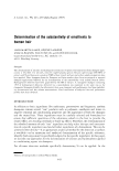
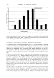
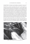
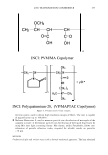
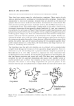
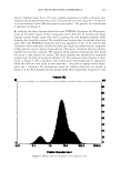
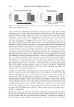
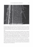
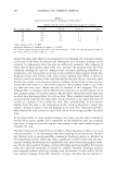
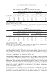
342 JOURNAL OF COSMETIC SCIENCE FLUORESCENCE MICROSCOPY The hair fibres were embedded into an epoxy resin and transversal cuts of the fibres with 10 µm were prepared using a microtome. Fibres cross-sections were analyzed by a transmission optic microscope (Olympus BH2) with a magnification of 40x. RES UL TS AND DISCUSSION The hair surface tends to be negatively charged at neutral and/or alkaline pHs. When hair is damaged, either by chemical or mechanical factors, its negative charge increases, increasing as well its friction and adhesion properties (7 ,8). In this particular study the peptides were formulated together with a lipid, phosphatidylcholine, which was added to attain a peptide formulation compatible with a water environment. Due to the large size of the Leucine side chain, the synthesized peptides tend to acquire an alpha helix structure in water. The interaction with phospholipids could further stabilize this alpha helix structure (17). However, the KAKAK sequence positioned at the C or N terminus, despite the large size of the amino acid Lysine, might induce a different structure for these two peptides, due to both charge and interaction with the hydrophobic parts of phospholipids. However, we do not know for sure the exact structure of these peptides in water. The peptide sequences were visualized by a molecular modelling program to identify the major differences in their structure (Figure 1). The molecular modelling program allows for creating the structure based on the amino acids sequence, which only differs in the position of the charged group (KAKAK), which is at the C-terminus or at the N- terminus, respectively, in the C-term or N-term peptides. Figure 1 shows the structures of C-term and N-term peptides in vacuum. Besides illustrating the amphipathic nature of the helix, it also shows a much narrower spatial distribution of the positively charged side chains in the C-term peptide. These peptides tend to be therefore both amphipathic and cationic. Amphipathicity increases their affinity for biological membranes, while the positive charge increases their specificity toward negatively charged membranes, as those of hair ( 1 7, 18). The total net charge of these peptides was found to be +3. Accordingly to Sharadadevi et al. (19), helices with Figure 1. Surface charge analysis for the C-term (A) and N-term (B) peptides, from PyMol v0.99. Red denotes the negatively charged C-terminus while blue denotes the positively charged side chains.
Purchased for the exclusive use of nofirst nolast (unknown)
From: SCC Media Library & Resource Center (library.scconline.org)