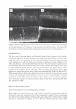
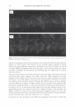
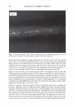
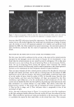
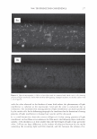
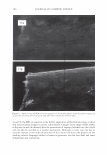
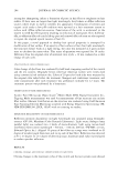
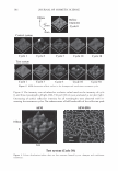
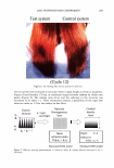
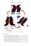
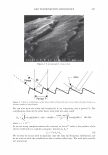
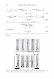
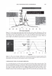
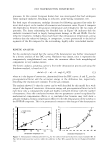
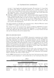
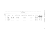
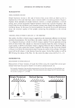
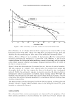
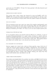
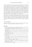
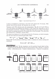
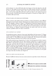
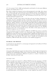
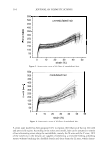
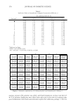
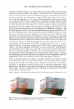
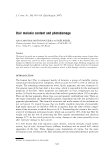
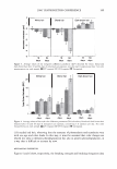
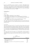
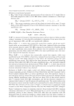
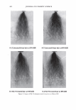
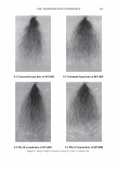
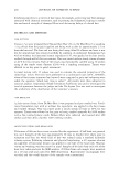
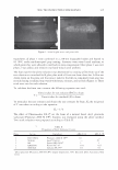
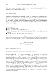
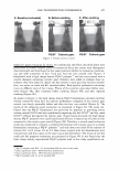
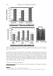
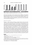
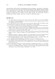
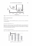
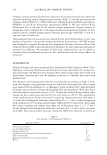
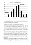
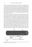
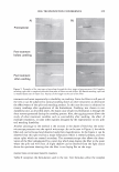
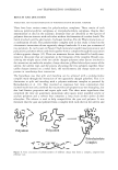
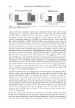
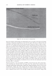
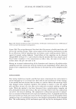
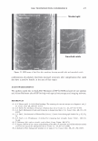
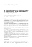
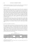
2006 TRI/PRINCETON CONFERENCE 273 Figure 4. Optical (4a) and SEM (46) micrographs (x370) showing letails of dot like patterns appearing in cuticle cells after drying the hair fiber with a hot iron surface at T = 90 C for 5 minutes. Lines and circles in captions help to identify same cuticle sheath areas in both micrographs. cuticle cells were damaged. For instance, the LIPs of hyperbolic shape were seen invari- ably associated to the folding of cuticle cells in buckles. Figures 2a, 26, 2c, and 2d show the juxtaposition of microscopic images displaying the same area of a hair surface with de-cemented and buckled cuticle cells, one obtained by optical microscopy and the other by SEM, respectively. In these figures it can be seen that each cuticle cell that appears folded forming buckles in the SEM micrograph (see Figures 2a and 26) has a corre- sponding hyperbolic pattern of light interference in the picture obtained by optical microscopy (see Figures 2c and 2d). The hyperbolic LIPs were found to be very sensltlve to the application of additional mechanical or swelling stresses to the cuticle cells. Their color and shape underwent dramatic changes when their corresponding cuticle cells were further stressed. Figures 6a and 66 show before and after pictures of a de-cemented and buckled cuticle cell that was

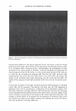
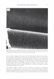
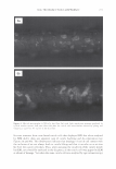
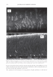
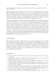
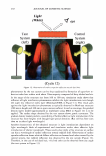
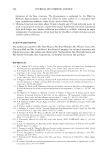
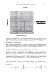
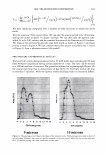
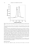
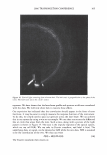
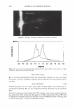
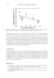
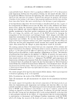
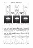
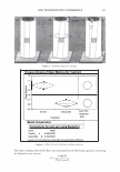
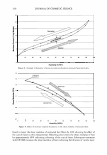
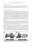
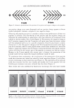
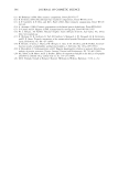
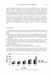
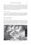
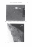
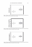
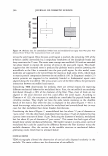
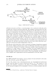
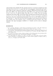
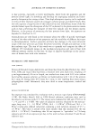
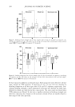
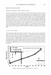
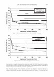
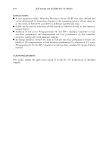
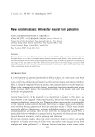
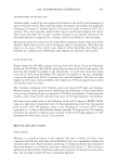
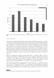
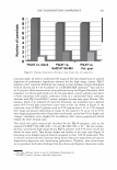
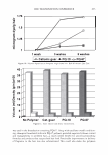
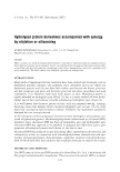
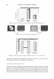
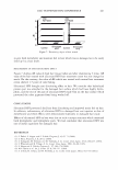
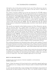
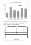
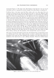
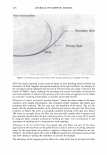
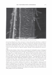
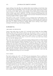
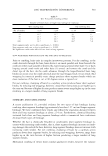
274 JOURNAL OF COSMETIC SCIENCE Figure 5. Optical micrograph (x250) of light interference pattern in channel form produced on hair by cyclical torsion stresses on a hair fiber using the protocol described elsewhere (13 ). further stressed by applying a slight pulling force to bend the cuticle cell. The pulling force to the cuticle cells was applied by gluing a tape to the hair surface and subsequently lifting the tape. The adhesion force of the tape was such that it didn't lead to cuticle breakage. The main change observed after application of the tape was the color and shape of the cuticle cell's LIP that changed from one displaying colored bands hyperbolic in nature to one of straight colored lines or wide bands (see Figures 6a and 66). Figures 7a and 76 show, on the other hand, the effects of isopropyl alcohol on the LIPs of de-cemented and buckled cuticle cells. These figures show that the color and shape of the LIPs has changed after the hair fiber was immersed in isopropyl alcohol for 1 minute. It was also observed that if a de-cemented and buckled cuticle cell was allowed to recover from its deformed state by immersing it in water its LIP disappeared. It is interesting to mention here that SEM analysis of cuticle cells recovered in this manner showed them apparently re-glued again to the hair surface. However, treatment of water re-glued cuticle cells with IPA caused them to buckle and lift again. Although, their new deformed shapes differed from those originally formed. Treatment of hair with IPA can be used, thus, to reveal breakage of cuticular cement that may have occurred previously and that has been concealed by a water recovery process. The treatment of virgin hair fibers taken from scalp close to the root with IPA never caused by itself cuticle cell de-cementation and buckling. The application of oils such as silicone and esters to de-cemented and buckled cuticle cells didn't lead to their me- chanical recovery, but instead it caused the suppression of the LIPs (see Figures Sa and 86).
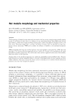
Purchased for the exclusive use of nofirst nolast (unknown)
From: SCC Media Library & Resource Center (library.scconline.org)