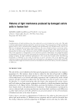
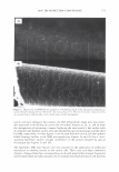
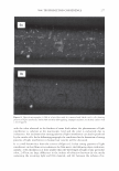
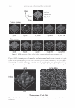
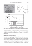
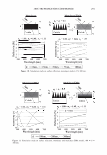
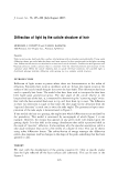
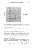
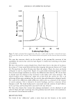
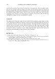
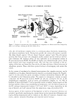
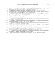
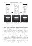
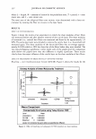
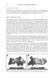
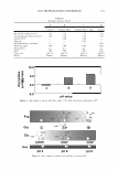
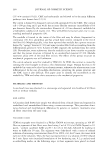
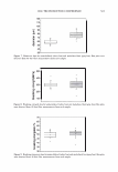
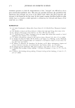
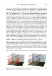
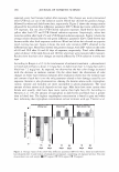
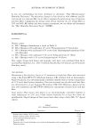
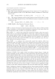
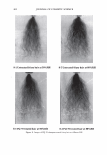
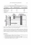
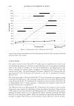
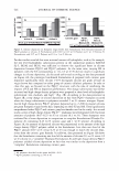
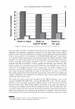
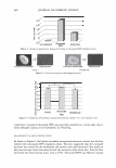
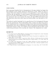
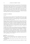
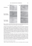
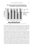
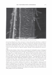
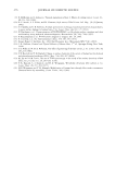
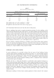
2006 TRI/PRINCETON CONFERENCE 275 Fi g ure 6. Optical micrograpbs (x2'50) of a hair fiber (6a) with light intcrfr-rence patterns produced hy cyclical tension stresses, and same fiber (66) after rhe curiclc cells were further stressed by gluing and srripping a rape from rlw surface of the hair fiber. In some instances there were found cuticle cells that dis played LIPs but when analyzed by SEM didn't show any apparent sign of cuticle buckling and de-cementation (see Figures 9a and 96). This observation indicates that breakage of cuticle cell cement with the inclusion of air not always leads to cuticle lifting and that it can also occur at sites far from the cuticle cell edges. Thus, when assessing the condition of the cuticle sheath by SEM, care should be exercised in the diagnosis, as the cuticle cell may appear by SEM as absent of damage. Yet when the same cuticle cells are analyzed by optical microscopy

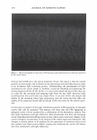
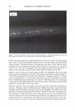
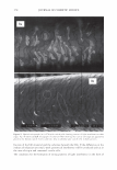
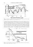
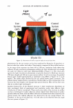
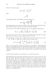
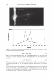
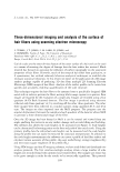
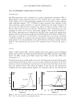
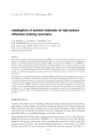
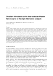
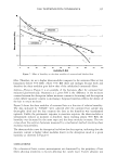
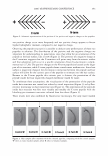
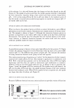
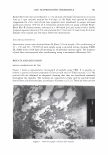
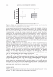
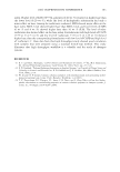
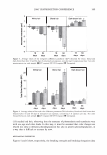
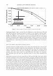
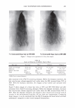
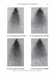
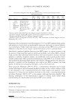
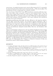
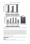
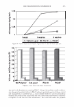
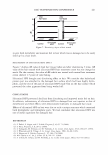
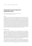
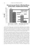
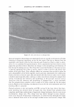
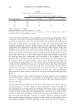
276 JOURNAL OF COSMETIC SCIENCE Figure 7. Optical micrographs (x300) of a hair fiber with de-cemented and lifted cuticle cells showing patterns of light interference before (7a) and after (76) immersion in isopropyl alcohol for 1 minute. they may show LIPs indicating internal de-cementation. The LIPs were also produced by various cuticle cells stacked together that underwent de-cementation in lumps. In many cases the extent of cuticle cell separation appeared to be shallow and superficial when analyzed by SEM, but when analyzed by optical microscopy, the associated pattern of light interference indicated that the extent of separation was deep inside the cuticle sheath. MECHANISM FOR THE FORMATION OF LIPS OF LINEAR AND HYPERBOLIC SHAPE The first issue that will be addressed in this section is the origin of the colored patterns displayed by the damaged cuticle cells shown in Figures 2a-2d. Incidentally, it was found that the colored patterns are not caused by stress bi-refringency ( 14) as they were observed to occur even without the use of polarizing filters in the microscope. Further- more, the use of polarizing filters during the analysis didn't eliminate the colored patterns at all. Instead, the filters only caused a small attenuation in the colored pattern's intensity so the explanation for their formation should be looked elsewhere. There are many examples of other colored patterns produced by the physical interaction of light with matter and these can be used as a starting basis for the analysis of LIPs observed on cuticle cells. For instance, the colors in soap bubbles, in Newton rings, from oil on the surface of water, from the surface of CDs, in butterfly wings, from the skin of some beetles, and from some bird feathers, all them are produced by thin film interference, light diffraction, or by the combination of both mechanisms (15, 16). The cuticle cells in human hair have also the capability of producing colored patterns by thin film interference in a similar fashion to a soap bubble. This ability stems from the following facts, namely: 1) The cuticle cells are composed of various transparent layers, 2) They are flat in shape, and 3) Their thickness value is comparable to that of the wavelength of light. In fact, the weak iridescence shown in Figure 1 is certainly due to thin film interference as the light undergoes double reflection from the cuticle cells near the surface of the hair fiber. These interference patterns are weak and incoherent at the macroscopic level, and it is probably the reason for their small effect on the overall hair color. This contrasts
Purchased for the exclusive use of nofirst nolast (unknown)
From: SCC Media Library & Resource Center (library.scconline.org)